Ki-67 image by IHC-12376 - Technical only, 12379 - Technical & Interpretation
Test info
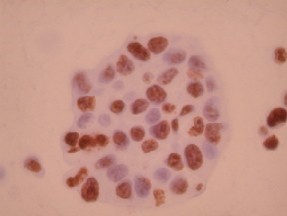
Ki-67 image by IHC
12376 - Technical only, 12379 - Technical & Interpretation
LAB12376
LAB12379
LAB12379
IHC
Mib-1
- All IHC stains will include a positive control tissue
- In paraffin embedded, formalin fixed tissue, the antibody labels cells that are cycling
- This antibody has been used as an index of proliferation to assess the biologic potential of various tumors
- Ki-67 is also useful in cervical biopsies, and can help differentiate between dysplasia (positive staining corresponding to the degree of dysplasia) and reactive atypia (positive staining only along basal epithelium)
Specimen
Tissue
Submit a formalin-fixed, paraffin embedded tissue
Formalin-fixed, paraffin embedded (FFPE) tissue block
FFPE tissue section mounted on a charged, unstained slide
Ambient (preferred)
- Unlabeled/mislabeled block
- Insufficient tissue
- Slides broken beyond repair
Performance
AHL - Immunohistochemistry
Mo - Fr
1 - 2 days
Immunohistochemical staining and microscopic examination
Clinical and Interpretive info
If requested, an interpretive report will be provided
Specifications
- The antibody reacts with a nuclear antigen which is expressed throughout the cell cycle (G1, S, G2, M phases)
- Ki-67 is never expressed by resting cells (in G0 phase)
Staining pattern
- Nuclear only
References
- Gerdes et al: Immunobiochemical and molecular biologic characterization of the cell proliferation-associated nuclear antigen that is defined by monoclonal antibody Ki-67. 1991, Am J Pathol, 4, 138, 8670873.
- Al-Saleh W et al: Assessment of Ki-67 Antigen Immunostaining in Squamous Intraepithelial Lesions of the Uterine Cervix. Am J Clin Pathol 1995:104:154-160, 1995.
Billing
88342 - 1st stain
88341 - each additional stain
88361 - CAS Morphometric
88341 - each additional stain
88361 - CAS Morphometric
Tracking
07/16/2017
02/25/2019
01/12/2024