STAT6 by IHC
STAT6 by IHC-12376 - Technical only, 12379 - Technical & interpretation
STAT6 by IHC
12376 - Technical only, 12379 - Technical & interpretation
LAB12376
LAB12379
LAB12379
- All IHC stains will include a positive control tissue
- SFT have a relatively unspecific IHC profile characterized by the co-expression of CD34, CD99, and BCL2. However, this profile is typically not seen in dedifferentiated tumors
- One recent study showed staining for STAT6 in 206 of 240 SFTs
- Of 1781 soft tissue tumors, STAT6 staining was seen in 4% of non SFT
- STAT6 staining has also been reported in 49 of 408 well-differentiated/de-differentiated liposarcomas, and eight of 65 unclassified sarcomas, and 14 of 184 desmoid tumors (but these tumors are generally histologically distinguishable from SFT)
- STAT6 is a sensitive and specific marker for identifying hemangiopericytoma/solitary fibrous tumor (HPC/SFT) of the brain and spinal cord. This marker can be used in conjunction with SSTR2a (a sensitive marker for meningiomas)
Tissue
Submit a formalin-fixed, paraffin embedded tissue block
Formalin-fixed, paraffin embedded (FFPE) tissue block
FFPE tissue section mounted on a charged, unstained slide
Ambient (preferred)
- Unlabeled/mislabeled block
- Insufficient tissue
- Slides broken beyond repair
AHL - Immunohistochemistry
Mo - Fr
1 - 2 days
Immunohistochemical staining and microscopic examination
If requested, an interpretive report will be provided
Specifications
- STAT6 is a highly sensitive and specific transcription factor for solitary fibrous tumors (SFT)
- Recently NAB2/STAT6 gene fusion has been recognized in almost all SFT, and can be identified in typical and malignant variants
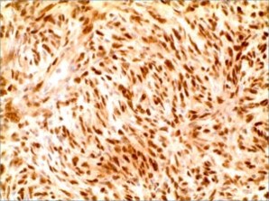
Staining pattern
- Strong diffuse nuclear staining is seen in usual and malignant SFT
- Dedifferentiated tumors may show negative or heterogeneous staining (these tumors may also show weak cytoplasmic staining)
- Caution should be utilized in meningiomas, since prominent cytoplasmic staining may be observed, and could be misinterpreted as nuclear staining
References
- Dagrada GP et al: Solitary fibrous tumors: loss of chimeric protein expression and genomic instability mark dedifferentiation. Modern Pathology (2015) 28, 1074-1083; doi:10.1038/modpathol.2015.70.
- Demicco DG et al: Extensive Survey of STAT6 Expression in a Large Series of Mesenchymal Tumors. Am J Clin Pathol 2015;143:672-682.
- Doyle LA et al: Nuclear expression of STAT6 distinguishes solitary fibrous tumor from histologic mimics. Modern Pathology (2014) 27, 390-395.
- Koelsche C et al: Nuclear relocation of STAT6 reliably predicts NAB2-STAT6 fusion for the diagnosis of solitary fibrous tumour. Histopathology 2014, 65, 613-622. DOI: 10.1111/his.12431.
- Yoshida A et al: STAT6 Immunohistochemistry Is Helpful in the Diagnosis of Solitary Fibrous Tumors. Am J Surg Pathol 2014;38:552-559.
- Schweizer L et al: Meningeal hemangiopericytoma and solitary fibrous tumors carry the NAB2-STAT6 fusion and can be diagnosed by nuclear expression of STAT6 protein. Acta Neuropatholo. 2013;125(5)651-8.
88342 - 1st stain
88341 - each additional stain
88341 - each additional stain
09/13/2017
10/17/2018
01/12/2024