MYB by IHC
MYB by IHC-12376 - Technical only, 12379 - Technical & interpretation
MYB by IHC
12376 - Technical only, 12379 - Technical & interpretation
LAB12376
LAB12379
LAB12379
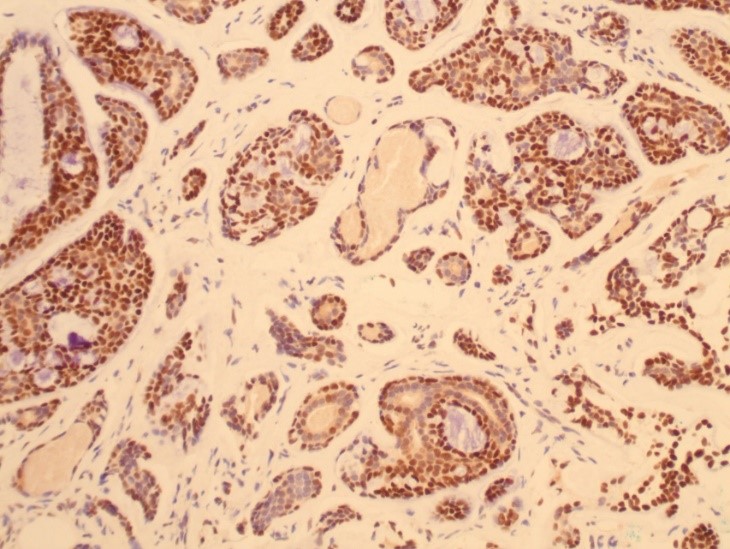
MYB
- All IHC stains will include a positive control tissue
The MYB antibody is useful identifying adenoid cystic carcinoma arising in the breast
Tissue
Submit a formalin-fixed, paraffin embedded tissue block
Formalin-fixed, paraffin embedded (FFPE) tissue block
FFPE tissue section mounted on a charged, unstained slide
Ambient (preferred)
- Unlabeled/mislabeled block
- Insufficient tissue
- Slides broken beyond repair
AHL - Immunohistochemistry
Mo - Fr
1 - 2 days
Immunohistochemical staining and microscopic examination
If requested, an interpretive report will be provided
Specifications
- A recurrent translocation is found in subset of adenoid cystic carcinomas (ACC), (t(6;9)(q22-23;p23-24)) resulting in the fusion of the oncogene MYB with the transcription factor NFIB.
- The MYB-NFIB fusion gene results in increased MYB protein overexpression by IHC.
- This antibody is useful identifying adenoid cystic carcinoma arising in the breast.
Staining pattern
- Nuclear (diffuse, moderate to strong staining); this staining pattern can be confined to the myoepithelial cells.
- In resection specimens, this antibody can demonstrate a peripheral staining pattern, presumably due to slower formal fixation and short half-life of the MYB protein.
- Weak or focal staining can be seen other entities arising in the breast (including collagenous spherulosis, basal-like triple negative breast cancers).
- Weak or focal staining can be seen other entities arising in the breast (including collagenous spherulosis, basal-like triple negative breast cancers).
Applications:
- Breast ACC: Diffuse, moderate to strong MYB IHC staining; sensitivity of 100%, specificity of 100%.
- Focal, weak MYB IHC staining has been described in triple negative breast carcinoma and collagenous spherulosis
- The MYB IHC stain is more sensitive and specific than FISH testing in identifying ACC of the breast. MYB FISH is reported to be 96% sensitive and 89% specific, but 46% of MYB-NFIB negative ACC by FISH testing will stain for MYB IHC
- Salivary gland ACC: MYB IHC sensitivity of 82%, specificity of 86%.
- Cutaneous ACC: Small case series suggests high sensitivity (8 of 9 cases positive)3
- MYB IHC positivity has been described in DCIS, non- adenoid cystic salivary gland neoplasms (acinic cell carcinoma, basal cell adenoma, basal cell adenocarcinoma, epithelial-myoepithelial carcinoma, monomorphic carcinoma, mucoepidermoid carcinoma, pleomorphic adenoma, polymorphous low-grade adenocarcinoma, salivary duct carcinoma, salivary adenoma NOS), squamous cell carcinoma, basaloid squamous cell carcinoma, lymphoma, thymoma, colonic adenocarcinoma, melanoma, skin adnexal tumors (dermal cylindromas and eccrine spiroadenomas), sinonasal rhabdomyosarcoma, and nasopharyngeal carcinoma.
References:
- Xu B et al. Predictors of outcome in adenoid cystic carcinoma of salivary glands: a clinicopathologic study with correlation between MYB fusion and protein expression Am J Surg Pathol 2017:41(10):1422-1432.
- Poling JS et al: MYB Labeling by Immunohistochemistry Is More Sensitive and Specific for Breast Adenoid Cystic Carcinoma than MYB Labeling by FISH, Am J Surg Pathol 2017;41:973–97.
- North JP et al. Detection of MYB alterations and other immunohistochemical markers in primary cutaneous adenoid cystic carcinoma. Am J Surg Pathol. 2015;39: 1347–1356.
- Brill LB 2nd, et al. Analysis of MYB expression and MYB-NFIB gene fusions in adenoid cystic carcinoma and other salivary neoplasms. Mod Pathol. 2011;24:1169–1176.
88342 - 1st stain
88341 - each additional stain
88341 - each additional stain
02/14/2020
01/12/2024