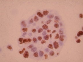
Ki-67 image by IHC
Ki-67 image by IHC-12376 - Technical only, 12379 - Technical & Interpretation
Ki-67 image by IHC
12376 - Technical only, 12379 - Technical & Interpretation
LAB12376
LAB12379
LAB12379
IHC
Mib-1
- All IHC stains will include a positive control tissue
- In paraffin embedded, formalin fixed tissue, the antibody labels cells that are cycling
- This antibody has been used as an index of proliferation to assess the biologic potential of various tumors
- Ki-67 is also useful in cervical biopsies, and can help differentiate between dysplasia (positive staining corresponding to the degree of dysplasia) and reactive atypia (positive staining only along basal epithelium)
Tissue
Submit a formalin-fixed, paraffin embedded tissue
Formalin-fixed, paraffin embedded (FFPE) tissue block
FFPE tissue section mounted on a charged, unstained slide
Ambient (preferred)
- Unlabeled/mislabeled block
- Insufficient tissue
- Slides broken beyond repair
AHL - Immunohistochemistry
Mo - Fr
1 - 2 days
Immunohistochemical staining and microscopic examination
If requested, an interpretive report will be provided
Specifications
- The antibody reacts with a nuclear antigen which is expressed throughout the cell cycle (G1, S, G2, M phases)
- Ki-67 is never expressed by resting cells (in G0 phase)
Staining pattern
- Nuclear only
References
- Gerdes et al: Immunobiochemical and molecular biologic characterization of the cell proliferation-associated nuclear antigen that is defined by monoclonal antibody Ki-67. 1991, Am J Pathol, 4, 138, 8670873.
- Al-Saleh W et al: Assessment of Ki-67 Antigen Immunostaining in Squamous Intraepithelial Lesions of the Uterine Cervix. Am J Clin Pathol 1995:104:154-160, 1995.
88342 - 1st stain
88341 - each additional stain
88361 - CAS Morphometric
88341 - each additional stain
88361 - CAS Morphometric
07/16/2017
02/25/2019
01/12/2024