HER-2/neu by IHC
HER-2/neu by IHC-12376 - Technical only, 12379 - Technical & interpretation
HER-2/neu by IHC
12376 - Technical only, 12379 - Technical & interpretation
LAB12376
LAB12379
LAB12379
c-erB-2
- All IHC stains will include a positive control tissue
Breast Carcinoma
- HER-2/neu (c-erbB-2) oncoprotein is a 185 kd protein with structural similarities to epidermal growth factor receptor (EGFR). From 25-40% of breast cancers over-express HER-2/neu protein. It's over-expression in breast cancer is associated with high risk of early relapse and poor prognosis, regardless of estrogen receptor status
- In axillary node-positive patients, HER-2/neu over-expression is associated with shorter disease-free and overall survival. In node-negative patients, HER-2/neu over-expression correlates with decreased survival only among women with tumors showing good nuclear grade compared with those negative for HER-2/neu staging (five times higher mortality rate, 0.117 versus 0.024 respectively). This may have value in distinguishing patients at increased risk within a relatively low-risk group, who may benefit from adjuvant therapy and close clinical monitoring
- A primary tumor may be HER-2/neu negative but subsequent metastases may express HER-2/neu protein. However, if a primary tumor expresses HER-2/neu, the positivity is retained in all metastases
- Regarding ductal in-situ carcinomas, multiple studies show HER-2/neu staging to correlate with increased nuclear size, 15-20 um (94% immunoreactivity versus 0% for small nuclei, <10% um), and with type of DCIS (>50% positivity in comedo type, but almost never in cribriform or micropapillary patterns) 8
Ovarian Carcinoma
- HER-2/neu over-expression in epithelial ovarian cancer may be a marker of more advanced stage, worse response to chemotherapy, and shorter survival time. 9 These patients may respond to a more aggressive course of chemotherapy
Neuroblastoma
- HER-2/neu positivity in neuroblastoma denotes shorter survival time (median survival 13 months versus 138 months for HER-2/neu-negative tumors) 10
- The presence of HER-2/neu oncoprotein may be a useful indicator of prognosis in human carcinomas of the breast, uterus, ovary and gastrointestinal tract
Tissue
Submit a formalin-fixed, paraffin-embedded tissue
Formalin-fixed, paraffin-embedded (FFPE) tissue block
FFPE tissue section mounted on a charged, unstained slide
Ambient (preferred)
- Unlabeled/mislabeled block
- Insufficient tissue
- Slides broken beyond repair
AHL - Immunohistochemistry
Mo - Fr
1 - 2 days
Immunohistochemical staining and microscopic examination
If requested, an interpretive report will be provided
Specifications
- Reacts with HER-2/neu oncoprotein in formalin-fixed or Bouin's-fixed, paraffin-embedded tissue
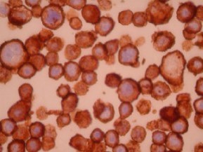
Staining pattern
- Strong cell membrane staining is shown to be associated with poor overall survival rate. (Cytoplasmic staining occurs and has been shown to be of no prognostic significance. Therefore, it is recommended that cytoplasmic staining be disregarded in the interpretation of HER-2/neu over-expression 5)
References
- Slamon DJ, Clark GM, Wong SG, Levin WJ, Ulrich A, McGuire WL. Human breast cancer: correlation of relapse and survival with amplification of the HER-2/neu oncogene. Science 1987; 235:177-82.
- Wright C, Angus B, Nicholson S, et al. Expression of c-erbB-2 oncoprotein : a prognostic indicator in human breast cancer. Cancer Res 1989; 49:2087-90.
- Barnes DM. Breast cancer and a proto-oncogene c-erbB-2 is a reliable prognostic marker. Br Med J 1989: 299:1061-2.
- Marks JR, Humphrey PA, Wu K, Berry D, Bandarenko N, Kerns BM, Inglehart JD. Overexpression of p53 and HER-2/neu proteins as prognostic markers in early stage breast cancer. Annals of Surgery 1994; 219(4):332-341.
- Tetu B, Brisson J. Prognostic significance of HER-2/neu oncoprotein expression in node-positive breast cancer: the influence of the pattern of immunostaining and adjuvant therapy. Cancer 1994; 73(9):2359-2365.
- DePotter CR. The neu-oncogene: more than a prognostic indicator? Human Pathology 1994; 25(12):1264-1268.
- Paik S, Hazan R, Fisher ER, Sass RE, Fisher B, Redmond C, Schlessinger J, Lippman ME, King ME, King CR. Pathologic findings from the national surgical adjuvant breast and bowel project: prognostic significance of erbB-2 protein over-expression in primary breast cancer. Journal of Clinical Oncology 1990; 8(1): 103-112.
- Bartkova J, Barnes DM, Millis RR, Gullick WJ. Immunohistochemical demonstration of c-erbB-2 protein in mammary ductal carcinoma in situ. Human Pathology 1990; 21(11):1164-1167.
- Felip E, Del Campo JM, Rubio D, Vidal MT, Colomer R, Bermejo B. Over-expression of C-erbB-2 in epithelial ovarian cancer: prognostic value and relationship with response to chemotherapy. Cancer 1995; 75(8):2147-2152.
- Layfield LJ, Thompson JK, Dodge RK, Kerns B. Prognostic indicators for neuroblastoma: stage, grade, DNA ploidy, MIB-1 proliferation index, p53, HER-2/neu and EGFr - a survival study. Journal of Surgical Oncology 1995; 59:21-27.
88342 - 1st stain
88341 - each additional stain
88341 - each additional stain
07/03/2017
10/19/2018
01/12/2024