ERG by IHC
ERG by IHC-12376 - Technical only, 12379 - Technical & interpretation
ERG by IHC
12376 - Technical only, 12379 - Technical & interpretation
LAB12376
LAB12379
LAB12379
ETS-related gene
All IHC stains will include a positive control tissue
Applications:
- ERG is a sensitive and specific marker of endothelial differentiation, and is more specific than CD31, CD34 and FLI1. Thus, this is a useful marker for benign and malignant vascular tumors
- ERG is not present in pericytes. Thus, the presence of a dual cell population, ERG positive endothelial cells and ERG negative nonendothelial components can be helpful in favoring highly cellular hemangiomas over angiosarcoma (which would only have an ERG positive endothelial component)
- ERG has been reported in other vascular lesions such as littoral cell angioma (LCA). It is negative in peliosis
- Approximately 50% of 50% of prostate cancers are ERG positive, specifically including prostate carcinomas with TMPRSS2-ERG gene fusions that lead to the overexpression of ERG
- In bone marrow, it can be expressed in myeloid precursors (marrow stem cells are known to express ERG)
- Focal reactivity has been reported in rare undifferentiated lung tumors and mesotheliomas
Tissue
Submit a formalin-fixed, paraffin-embedded tissue
Formalin-fixed, paraffin-embedded (FFPE) tissue block
FFPE tissue section mounted on a charged, unstained slide
Ambient (preferred)
- Unlabeled/mislabeled block
- Insufficient tissue
- Slides broken beyond repair
AHL - Immunohistochemistry
Mo - Fr
1 - 2 days
Immunohistochemical staining and microscopic examination
If requested, an interpretive report will be provided
Specifications
- This antibody detects wildtype ERG and truncated ERG (resulting from gene rearrangement)
- ERG is a transcription factor expressed in vessel endothelium
- ERG is also expressed in a subset of prostate carcinomas
- This marker is also expressed in acute myeloid leukemia and Ewing sarcoma
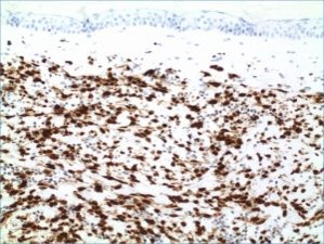
Staining pattern
- Nuclear staining
References
- O'Malley DP: Distinctive immunohistochemical staining in littoral cell angioma using ERG and WT-1. Ann Diagn Pathol. 2015 Mar 5. pii: S1092-9134(15)00041-6. doi: 10.1016/j.anndiagpath.2015.02.007. [Epub ahead of print].
- Shah RB et al: Heterogeneity of PTEN and ERG expression in prostate cancer on core needle biopsies: implications for cancer risk stratification and biomarker sampling. Hum Pathol. 2015 Jan 29. pii: S0046-8177(15)00028-3. doi: 10.1016/j.humpath.2015.01.008. [Epub ahead of print].
- Sullivan HC et al: The utility of ERG, CD31 and CD34 in the cytological diagnosis of angiosarcoma: an analysis of 25 cases. J Clin Pathol. 2015 Jan;68(1):44-50. doi: 10.1136/jclinpath-2014-202629. Epub 2014 Oct 28.
- Miettinen M et al: ERG Transcription Factor as an Immunohistochemical Marker for Vascular Endothelial Tumors and Prostatic Carcinoma. Am J Surg Pathol 2011;35:432-441).
88342 - 1st stain
88341 - each additional stain
88341 - each additional stain
06/21/2017
10/17/2018
01/12/2024