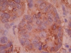
MART-1 by IHC
MART-1 by IHC-12376 - Technical only, 12379 - Technical & Interpretation
MART-1 by IHC
12376 - Technical only, 12379 - Technical & Interpretation
LAB12376
LAB12379
LAB12379
- All IHC stains will include a positive control tissue
- Expressed in virtually all nevi with the exception of neurotized melanocytes with prominent Schwannian differentiation
- Stains all melanomas with epithelioid or mixed spindle and epithelioid features
- Is more sensitive than HMB45 in identifying desmoplastic melanoma, however is less sensitive than S100 (Mart-1may stain <50% of cells in desmoplastic melanomas).
- Mart-1 can detect a small number of HMB45 negative metastatic melanomas and a rare case of S100 negative metastatic melanoma. It has virtually the same sensitivity as Melan-A; in one study, 81% of metastatic melanomas were reactive with Melan-A/Mart-1 and 75% with HMB45. 85% of cases stained for one or both antibodies
- Mart-1 has been proven to be a useful adjuvant to the HMB45/S100 panel for the diagnosis of malignant melanomas
- Mart-1 and HMB45 show similar staining in angiomyolipomas
- Mart-1 is less sensitive than HMB45 in identifying lymphangiomyomatosis (LAM), and clear cell tumors/sugar tumors (CCST) of lung; (LAM: 4/4 with HMB45 and ? with Mart-1; CCST of lung: 2/3 with HMB45 and 1/3 with Mart-1)
Tissue
Submit a formalin-fixed, paraffin embedded tissue
Formalin-fixed, paraffin embedded (FFPE) tissue block
FFPE tissue section mounted on a charged, unstained slide
Ambient (preferred)
- Unlabeled/mislabeled block
- Insufficient tissue
- Slides broken beyond repair
AHL - Immunohistochemistry
Mo - Fr
1 - 2 days
Immunohistochemical staining and microscopic examination
If requested, an interpretive report will be provided
Specifications
- Mart-1 is a transmembrane protein composed of 118 amino acids. It recognizes the same antigen as Melan-A (clone A103). However, Mart-1 does not cross react with steroid producing tumors (such as adrenocortical adenomas, primary and metastatic adrenocortical carcinomas, and Sertoli-Leydig cell tumors of the ovary and testes)
Staining pattern
- Cytoplasmic granular staining. The staining pattern is strong in epithelioid melanocytes
References
- Fetsch PA et al: The New Melanoma Markers: Mart-1 and Melan-A (The NIH Experience); Am J Surg Pathol, 23(5) 1999 607-609.
- Fetsch PA et al: Immunocytochemical Detection of Mart-1 in Fresh and Paraffin-embedded Malignant Melanoms; J Immunotherapy, 20(1) 1997, 60-64.
- Busam K et al: Expression of Melan-A (Mart-1) in Benign Melanocytic Nevi and Primary Cutaneous Malignant Melanoma. Am J Surg Pathol 22(8): 976-982, 1998.
- Jungbluth A et al: A103: An Anti-Melan-A Monoclonal Antibody for the Detection of Malignant Melanoma in Paraffin-embedded Tissues. Am J Surg Pathol 22(5): 595-602, 1998.
- Orchard G: Melan A(Mart-1); A new Monoclonal Antibody for Malignant Melanoma Diagnosis. Br J Biomed Sci 1998; 55: 8-15.
88342 - 1st stain
88341 - each additional stain
88341 - each additional stain
07/17/2017
01/31/2024
01/31/2022