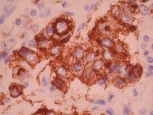
Prolactin by IHC-12376 - Technical only, 12379 - Technical & interpretation
Test info
LAB12379
- All IHC stains will include a positive control tissue
IHC staining for the presence of pituitary hormones helps distinguish pituitary adenomas and the rare primary pituitary carcinomas from other tumors occurring in the region of the sella (such as craniopharyngioma, metastatic carcinoma, meningioma, lymphoma, leukemia, and plasmacytoma), none of which contain or produce pituitary hormones Positive staining for specific pituitary hormones can divide the pituitary adenomas into subclasses based on the corresponding cell type present Negative staining for pituitary hormones does not rule out a pituitary adenoma since some contain none of the known adenohypophyseal hormones Prolactinomas often show a characteristic globular immunohistochemical reaction for PRL in the region of the paranuclear Golgi.
Specimen
Prepare a formalin-fixed, paraffin-embedded (FFPE) tissue block
Formalin-fixed, paraffin-embedded (FFPE) tissue block
Mount FFPE tissue section on a charged, unstained slide
Ambient (preferred)
- Unlabeled/mislabeled block
- Insufficient tissue
- Slides broken beyond repair
Performance
Immunohistochemical staining and microscopic examination
Clinical and Interpretive info
If requested, an interpretive report will be provided
Specifications
- This antibody reacts with prolactin hormone
- It recognizes the prolactin-secreting cells of the pituitary gland (also termed lactotrophs or mammotrophs)
- In the normal pituitary gland, these are mostly contained in the lateral wing of the adenohypophysis (posterolateral edges) and account for 15-25% of the cells of the gland
Staining pattern
- Cytoplasmic based staining
Billing
88341 - each additional stain

