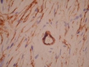
CD34 by IHC-12376 - Technical only, 12379 - Technical & interpretation
Test info
CD34 by IHC
12376 - Technical only, 12379 - Technical & interpretation
LAB12376
LAB12379
LAB12379
All IHC stains will include a positive control tissue
- 30-60% of B-ALL and AML's are positive for CD34
- CD34 is a sensitive, but nonspecific marker of vascular neoplasms
- CD34 has been shown useful in differentiating dermatofibroma (DF) from DFSP. CD34 is positive in the majority of cases of DFSP and negative in dermatofibromas
- CD34 has been identified in benign neural tumors such as neurofibromas, neuromas, and schwanomas1
- CD34 has been shown to be useful in differentiating solitary fibrous tumors of the pleura (SFT) from desmoplastic mesotheliomas. CD34 is positive in SFT and negative in mesotheliomas. Also included in the panel to differentiate these two lesions is Vimentin (positive in both) and CK (positive only in mesothelioma)2
- CD34 is useful in identifying gastrointestinal stromal tumors (GIST). Most GIST's may lack expression of all markers (including smooth muscle markers) except CD34
Specimen
Tissue
Submit a formalin-fixed, paraffin-embedded tissue
Formalin-fixed, paraffin-embedded (FFPE) tissue block
FFPE tissue section mounted on a charged, unstained slide
Ambient (preferred)
- Unlabeled/mislabeled block
- Insufficient tissue
- Slides broken beyond repair
Performance
AHL - Immunohistochemistry
Mo - Fr
1 - 2 days
Immunohistochemical staining and microscopic examination
Clinical and Interpretive info
If requested, an interpretive report will be provided
Specifications
- CD34 is a glycoprotein associated with human hematopoietic progenitor cells
- CD34 is present on immature hematopoietic precursor cells and all hematopoietic colony-forming cells in the bone marrow and blood
- CD34 is absent on fully differentiated hematopoietic cells
- In normal tissue, CD34 is present on endothelial cells, dermal dendritic cells, endoneurium of peripheral nerves and around dermal adnexal structures
Staining pattern
- Cytoplasmic
References
- Weiss et. al.; CD-34 Is Expressed by a Distinctive Cell Population in Peripheral Nerve, Nerve Sheath Tumors and Related Lesions; American Journal of Surgical Pathology 17(10): 1039-1045, 1993.
- Flint, et. al.; CD34 and Keratin Expression Distinguishes Solitary Fibrous Tumor (Fibrous Mesothelioma) of Pleura Form Desmoplastic Mesotheliomas; Human Pathol. 26:428-431, 1995
Billing
88342 - 1st stain
88341 - each additional stain
88341 - each additional stain
Tracking
06/16/2017
10/17/2018
01/12/2024
