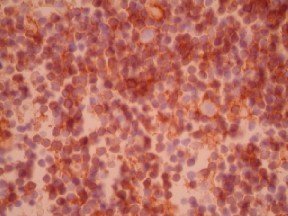
CD23 by IHC-12376 - Technical only, 12379 - Technical & interpretation
Test info
CD23 by IHC
12376 - Technical only, 12379 - Technical & interpretation
LAB12376
LAB12379
LAB12379
All IHC stains will include a positive control tissue
- CD23 is useful in a panel of immunostains (on frozen and/or paraffin sections) to differentiate between the various types of low grade B-cell lymphoma (CD23 is always positive in CLL/WDLL and only rarely and weakly positive in marginal zone lymphomas, follicular center cell lymphomas)
- CD23 was shown to stain Reed-Sternberg/Hodgkin’s cells in over 80% of cases in some series (nodular sclerosis type, LP, LD types). Approximately 15% of nodular sclerosing cases can be negative. In few cases CD23 was more useful than CD30, generally showing weaker staining. Therefore it should be used as part of a panel
- CD23 has been reported to be positive in rare cases of high grade T-cell lymphoma
Specimen
Tissue
Submit a formalin-fixed, paraffin-embedded tissue
Formalin-fixed, paraffin-embedded (FFPE) tissue block
FFPE tissue section mounted on a charged, unstained slide
Ambient (preferred)
- Unlabeled/mislabeled block
- Insufficient tissue
- Slides broken beyond repair
Performance
AHL - Immunohistochemistry
Mo - Fr
1 - 2 days
Immunohistochemical staining and microscopic examination
Clinical and Interpretive info
If requested, an interpretive report will be provided
Specifications
- CD23 is a 45 kD membrane glycoprotein identified as the low affinity IgE receptor (FcERII)
- CD23 reacts with activated B lymphocytes and some neoplastic B lymphocytes, dendritic reticulum cells, epidermal Langerhans cells, macrophages, some T lymphocytes, NK cells, Reed-Sternberg/Hodgkin’s cells, mast cells, occasionally endothelial cells
Staining pattern
- Cell membrane and diffuse cytoplasmic
- Intracellular (‘Golgi pattern’) in Reed-Sternberg/Hodgkin’s cells
References
- Rowlands et al. Journal of Pathology, 160:239-243, 1990.
- Murray et al. Journal of Pathology, 16J:125-128, 1991
Billing
88342 - 1st stain
88341 - each additional stain
88341 - each additional stain
Tracking
06/16/2017
10/17/2018
01/12/2024
