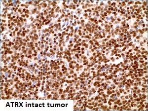
ATRX by IHC-12376 - Technical only, 12379 - Technical & interpretation
Test info
LAB12379
- All IHC stains will include a positive control tissue
ATRX Immunohistochemistry, Rationale and Clinical Significance:
The majority of grade II and III infiltrating gliomas and those glioblastomas derived from them have mutations of IDH1 codon 132 or IDH2 codon 172 7. Mutations of IDH1 or IDH2 are only very rarely found in other tumor types 8. An antibody to the most common mutant form of IDH1 (R132H), accounting for the vast majority of mutations detected, has recently been developed 9-11. Positive immunohistochemistry in this setting argues strongly for the diagnosis of infiltrating glioma 12, 13.
While IDH testing provides important information for the classification of infiltrating gliomas, additional subclassification has become the standard of care, since this provides additional important prognostic and predictive information6 and is incorporated into the 2016 update of the World Health Organization classification of tumors of the nervous system14.
Two large groups of IDH-mutant infiltrating gliomas are evident, and correspond roughly to the morphologic categories of oligodendroglioma (1p/19q codeleted) and astrocytoma (1p/19q nondeleted).
1p/19q codeletion is associated with oligodendroglial appearance, longer survival and better response to therapy, but its evaluation requires copy number analysis (often performed by fluorescence in situ hybridization), which is expensive and time consuming. Hence, in order to improve turnaround time, many laboratories rely on immunohistochemical stains for ATRX 15 and/or p53, which are associated with astrocytic differentiation.
Specimen
Submit a formalin-fixed, paraffin embedded tissue block
Formalin-fixed, paraffin embedded (FFPE) tissue block
Tissue section mounted on a charged, unstained slide
Ambient (preferred)
- Unlabeled/mislabeled
- Insufficient tissue
- Slides broken beyond repair
Performance
Immunohistochemical staining and microscopic examination
Clinical and Interpretive info
If requested, an interpretive report will be provided
Specifications
- Infiltrating gliomas are uncommon, incurable tumors of the brain and spinal cord
- Tumor cells variably resemble non-neoplastic astrocytes and oligodendroglia
- Comprehensive analysis of diffuse lower grade gliomas has shown that ATRX expression is lost in infiltrating gliomas that lack 1p/19q codeletion (1-6)
- In the setting of infiltrating glioma of grade II or III, ATRX immunohistochemistry is employed in most major neurosurgical centers as a means of avoiding 1p/19q analysis
- Loss of ATRX staining in neoplastic cells is strongly associated with astrocytic phenotype and 1p/19q nondeletion
Staining pattern
- Nuclear staining
References
- National Cancer Institute Genomic Data Commons Data Portal [cited 2017 08/06/2017]. Available from: https://portal.gdc.cancer.gov/.
- COSMIC Catalog of Somatic Mutations In Cancer [cited 2017 08/06/2017]. COSMIC, the Catalogue Of Somatic Mutations In Cancer, is the world's largest and most comprehensive resource for exploring the impact of somatic mutations in human cancer.]. Available from: http://cancer.sanger.ac.uk/cosmic.
- Tumor Portal [Available from: http://www.tumorportal.org/.
- FireBrowse [cited 2017 08/06/2017]. Available from: http://firebrowse.org/.
- Lawrence MS, Stojanov P, Mermel CH, Robinson JT, Garraway LA, Golub TR, et al. Discovery and saturation analysis of cancer genes across 21 tumour types. Nature. 2014;505(7484):495-501.
- Brat DJ, Verhaak RG, Aldape KD, Yung WK, Salama SR, Cooper LA, et al. Comprehensive, Integrative Genomic Analysis of Diffuse Lower-Grade Gliomas. N Engl J Med. 2015;372(26):2481-98. doi: 10.1056/NEJMoa1402121. Epub 2015 Jun 10.
- Yan H, Parsons DW, Jin G, McLendon R, Rasheed BA, Yuan W, et al. IDH1 and IDH2 mutations in gliomas. The New England journal of medicine. 2009;360(8):765-73.
- Gross S, Cairns RA, Minden MD, Driggers EM, Bittinger MA, Jang HG, et al. Cancer-associated metabolite 2-hydroxyglutarate accumulates in acute myelogenous leukemia with isocitrate dehydrogenase 1 and 2 mutations. The Journal of experimental medicine. 2010;207(2):339-44.
- Capper D, Sahm F, Hartmann C, Meyermann R, von Deimling A, Schittenhelm J. Application of mutant IDH1 antibody to differentiate diffuse glioma from nonneoplastic central nervous system lesions and therapy-induced changes. The American Journal of Surgical Pathology. 2010;34(8):1199-204.
- Capper D, Weissert S, Balss J, Habel A, Meyer J, Jager D, et al. Characterization of R132H mutation-specific IDH1 antibody binding in brain tumors. Brain pathology (Zurich, Switzerland). 2010;20(1):245-54.
- Capper D, Zentgraf H, Balss J, Hartmann C, von Deimling A. Monoclonal antibody specific for IDH1 R132H mutation. Acta Neuropathologica. 2009;118(5):599-601.
- Horbinski C, Kofler J, Yeaney G, Camelo-Piragua S, Venneti S, Louis DN, et al. Isocitrate dehydrogenase 1 analysis differentiates gangliogliomas from infiltrative gliomas. Brain pathology (Zurich, Switzerland). 2011;21(5):564-74.
- Preusser M, Wohrer A, Stary S, Hoftberger R, Streubel B, Hainfellner JA. Value and limitations of immunohistochemistry and gene sequencing for detection of the IDH1-R132H mutation in diffuse glioma biopsy specimens. Journal of neuropathology and experimental neurology. 2011;70(8):715-23.
- Louis DN, Ohgaki H, Wiestler OD, W.K. C, editors. World Health Organization Histological Classification of Tumours of the Central Nervous System: International Agency for Research on Cancer, France; 2016.
- Reuss DE, Sahm F, Schrimpf D, Wiestler B, Capper D, Koelsche C, et al. ATRX and IDH1-R132H immunohistochemistry with subsequent copy number analysis and IDH sequencing as a basis for an "integrated" diagnostic approach for adult astrocytoma, oligodendroglioma and glioblastoma. Acta Neuropathol. 2015;129(1):133-46. doi: 10.1007/s00401-014-1370-3. Epub 2014 Nov 27.
Billing
88341 - each additional stain
