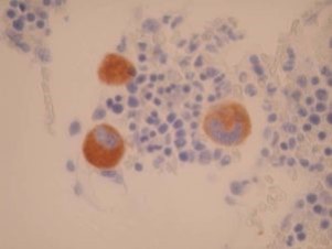
CD61 by IHC-12376 - Technical only, 12379 - Technical & interpretation
Test info
CD61 by IHC
12376 - Technical only, 12379 - Technical & interpretation
LAB12376
LAB12379
LAB12379
Platelet glycoprotein IIIa
All IHC stains will include a positive control tissue
- CD61 is the most sensitive and most specific of the platelet glycoprotein antibodies
- CD61 is the earliest glycoprotein to be expressed during normal megakaryocyte maturation
- 80% of AML-M7 react with CD61; 20% are more immature and lack CD61 (diagnosed by the presence of platelet peroxidase by EM)
- The platelets from patients with Glanzmann's thrombasthenia lack both glycoproteins IIb and IIIa
- CD61 can identify small platelet clumps (microthrombi) and may be of use in renal transplantation biopsies as well as studies of ischemic heart disease, inflammatory/ ischemic bowel disease, and placental pathology, etc.
- No cross reactivity with a variety of formalin-fixed paraffin-embedded normal and neoplastic tissues have been reported
Specimen
Tissue
Submit a formalin-fixed, paraffin-embedded tissue
Formalin-fixed, paraffin-embedded (FFPE) tissue block
FFPE tissue section mounted on a charged, unstained slide
Ambient (preferred)
- Unlabeled/mislabeled block
- Insufficient tissue
- Slides broken beyond repair
Performance
AHL - Immunohistochemistry
Mo - Fr
1 - 2 days
Immunohistochemical staining and microscopic examination
Clinical and Interpretive info
If requested, an interpretive report will be provided
Specifications
- CD61 is the integrin B-3-chain of the vitronectin receptor. This protein is found on platelets and megakaryocytes, and can associate with either glycoprotein IIb or vitronectin receptor ?-chain (CD51)
Staining pattern
- Cytoplasm of megakaryocytes, surface of platelets
- Well preserved in formalin
- Loss of antigenicity has been reported with mercuric fixatives
References
- Gatter KC, et al. The immunohistological detection of platelets, megakaryocytes and thrombi in routinely processed specimens. Histopathology 1988; 13:257-67.
- Erber WN, et al. Detection of cells of megakaryocyte lineage in hematological malignancies by immuno-alkaline phosphatase labeling cell smears with a panel of monoclonal antibodies. British Journal of Hematology 1987; 65:87-94.
- Cheson BD, et al. Report of the National Cancer Institute sponsored workshop on definitions of diagnosis and response in acute myeloid leukemia. Journal of Clinical Oncology 1990; 8(5):813-9.
Billing
88342 - 1st stain
88341 - each additional stain
88341 - each additional stain
Tracking
06/16/2017
10/18/2018
01/21/2026
