CD1a by IHC-12376 - Technical only, 12379 - Technical & interpretation
Test info
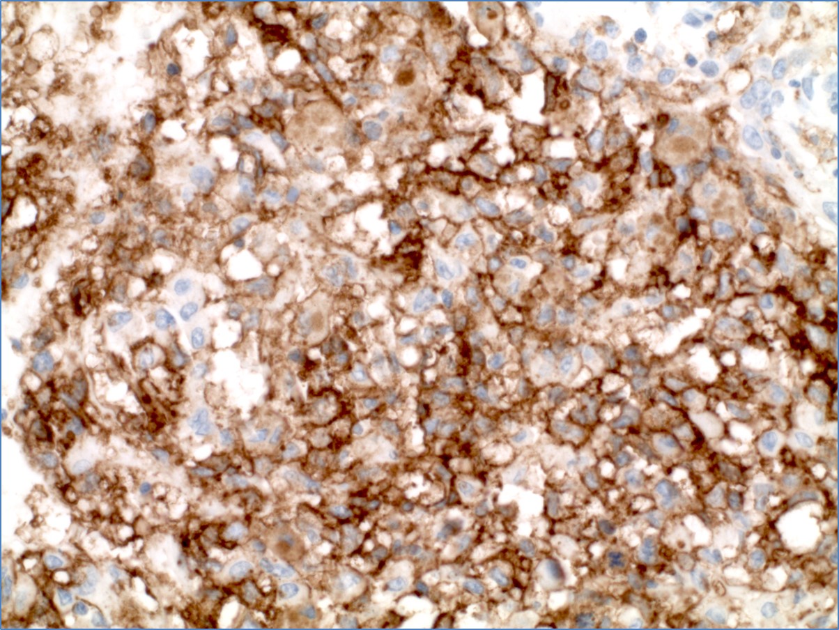
CD1a by IHC
12376 - Technical only, 12379 - Technical & interpretation
LAB12376
LAB12379
LAB12379
Leu-6
OKT6
SKg
T6
OKT6
SKg
T6
All IHC stains will include a positive control tissue
- CD1a can be used in conjunction with S-100 to differentiate between the various malignant and benign histiocytic proliferations:
| CD1a | S-100 | |
| Langerhans cells (LC)* | + | + |
| Interdigitating cells | - | + |
| Dendritic Reticulum cells | - | + |
* This pattern of staining is seen in both benign (e.g. dermatopathic lymphadenopathy) and malignant (histiocytic) LC proliferations. However, LC's in histiocytosis X have been found to be CD4 positive, while benign LC's are CD4 negative.
Specimen
Tissue
Submit a formalin-fixed, paraffin-embedded tissue
Formalin-fixed, paraffin-embedded (FFPE) tissue block
FFPE tissue section mounted on a charged, unstained slide
Ambient (preferred)
- Unlabeled/mislabeled block
- Insufficient tissue
- Slides broken beyond repair
Performance
AHL - Immunohistochemistry
Mo - Fr
1 - 2 days
Immunohistochemical staining and microscopic examination
Clinical and Interpretive info
If requested, an interpretive report will be provided
Specifications
- CD1a is a 49kDa cell surface glycoprotein found on human Langerhans cells and thymocytes, but not on peripheral T-cells
Staining pattern
- Cell membrane and/or diffuse cytoplasmic
References
- Ornvold et al, Virchows Archiv-A, 416(5):403-10, 1990.
- Shamoto et al, Advances in Exp. Med & Biology, 378:139-41, 1995.
Billing
88342 - 1st stain
88341 - each additional stain
88341 - each additional stain
Tracking
05/15/2017
10/17/2018
01/20/2026