TCL-1 by IHC-12376 - Technical only, 12379 - Technical & interpretation
Test info
TCL-1 by IHC
12376 - Technical only, 12379 - Technical & interpretation
LAB12376
LAB12379
LAB12379
- All IHC stains will include a positive control tissue
- TCL1 identifies blastic plasmacytoid dendritic cell neoplasm (BPDCN), which is a rare, highly malignant tumor with strong cutaneous tropism (nearly 100%) and leukemic phase either present at initial diagnosis (approximately 50% of cases) or in the subsequent course of the disease. Approximately 10-20% of cases are associated with or develop into AML
- TCL1 should be used in an antibody panel to detect BPDCN, which are CD4+, CD56+, CD123+ and TCL1+. CD68 is positive in approximately 50% of cases (often cytoplasmic dot staining). TdT is positive in approximately 20-60% of cases. Suspected cases of BPDCN should be subjected to rigorous immunophenotyping (including MPO and flow cytometry, if available), to assess for acute leukemia
- When only CD4, CD56 and CD123 are positive and TCL1 is negative, AML should be strongly considered
- Tumors that share some but not all immunophenotypic features of BPDCN are better classified as "acute leukemia of ambiguous lineage"
- TCL1 is also positive in most cases (75%) of T-PLL
Specimen
Tissue
Submit a formalin-fixed, paraffin embedded tissue block
Formalin-fixed, paraffin embedded (FFPE) tissue block
FFPE tissue section mounted on a charged, unstained slide
Ambient (preferred)
- Unlabeled/mislabeled block
- Insufficient tissue
- Slides broken beyond repair
Performance
AHL - Immunohistochemistry
Mo - Fr
1 - 2 days
Immunohistochemical staining and microscopic examination
Clinical and Interpretive info
If requested, an interpretive report will be provided
Specifications
- TCL1 (T-cell leukemia/lymphoma protein 1A) was originally identified in association with T-cell prolymphocytic leukemia (T-PLL). TCL1 reacts with human TCL1, which is involved with AKT phosphorylation/activation and is thought to enhance cell proliferation, stabilize mitochondrial membrane potential and promote cell survival
- TCL1 is normally expressed in immature cortical thymocytes, activated peripheral T-cells, pro-B cells, and naïve mantle zone B-cells of peripheral lymphoid tissues, but not in post-germinal center B-cells and plasma cells
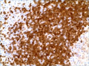
Staining pattern
- Nuclear and cytoplasmic staining
References
- WHO (2008) classification of tumors of hematopoietic and lymphoid tissues, p145-147.
- Am J Surg Pathol 2014;38:673-680
Billing
88342 - 1st stain
88341 - each additional stain
88341 - each additional stain
Tracking
09/13/2017
10/17/2018
01/12/2024