Mycobacterium tuberculosis by IHC-12376 - Technical only, 12379 - Technical & interpretation
Test info
Mycobacterium tuberculosis by IHC
12376 - Technical only, 12379 - Technical & interpretation
LAB12376
LAB12379
LAB12379
TB
- All IHC stains will include a positive control tissue
- The primary use for this antibody is in the detection of mycobacterial antigens in tissues and smears; this technique is not, however, specific for different species of mycobacteria
- A major benefit of this immunohistochemical technique as opposed to the Fite stain is that intact organisms need not be present; fragmented organisms, mycobacterial debris and organisms whose antigenicity may have been altered by antimycobacterial therapy will also be detected
- Minimal cross-reactivity has been reported (see "Staining Pattern" above).
Specimen
Tissue
Submit a formalin-fixed, paraffin embedded tissue block
Formalin-fixed, paraffin embedded (FFPE) tissue block
FFPE tissue section mounted on a charged, unstained slide
Ambient (preferred)
- Unlabeled/mislabeled block
- Insufficient tissue
- Slides broken beyond repair
Performance
AHL - Immunohistochemistry
Mo - Fr
1 - 2 days
Immunohistochemical staining and microscopic examination
Clinical and Interpretive info
If requested, an interpretive report will be provided
Specifications
- Reacts with approximately 100 different BCG antigens, many of which are common to mycobacteria other than M. bovis
- A few of these antigens are common to bacteria from families other than Mycobacterium (see below)
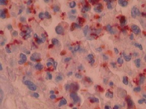
Staining pattern
- In contrast to the Fite stain, this antibody stains not only entire, intact organisms but also mycobacterial fragments and debris
- Because this antibody stains debris and fragmented organisms, staining can be identified in necrotic, caseous areas (unlike the Fite stain)
- Staining has been identified in typical caseating granulomas as well as in atypical granulomatous lesions
- Cross-reactivity with Corynebacterium spp., Nocardia spp., Rhodococcus spp., sporotrichosis and fungal organisms has been reported. However, these organisms may be distinguishable morphologically
References
- Hove MGM, Smith MB, et al. Detection of mycobacteria with use of immunohistochemistry in granulomatous lesions staining negative with routine acid-fast stains. Appl Immunohistochem 6(3): 169-72, 1998.
- Carabias E, Palenque E, et al. Evaluation of an immunohistochemical test with polyclonal antibodies raised against mycobacteria used in formal-fixed tissue compared with mycobacterial specific culture. APMIS 106: 385-8, 1998.
- Data sheet: DAKO.
Billing
88342 - 1st stain
88341 - each additional stain
88341 - each additional stain
Tracking
07/18/2017
10/19/2018
01/12/2024