p40 by IHC-12376 - Technical only, 12379 - Technical & interpretation
Test info
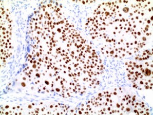
p40 by IHC
12376 - Technical only, 12379 - Technical & interpretation
LAB12376
LAB12379
LAB12379
- All IHC stains will include a positive control tissue
- p40 is equivalent to p63 in sensitivity for lung SCC, but is markedly more specific
- p40 stains fewer lung adenocarcinoma cases compared to p63; 0.8% of lung adenocarcinomas stained in one study 1
- Most urothelial cancers are p40 positive (85%)
- 95% of skin SCC and basal cell carcinomas are p40 positive
- p40 is equivalent to p63 in staining myoepithelial cells of breast and prostate
- Recent study showed p40-positive cases were as follows: 153 (96.8%) of 158 SCCs, 7 (4.6%) of 152 adenocarcinomas, 0 (0%) of 50 carcinoid tumors, 4 (3.6%) of 107 large cell neuroendocrine carcinomas, 1 (1.5%) of 68 small cell lung carcinomas, and 1 (2.4%) of 41 mesotheliomas2
- p40 is a useful marker for excluding neuroendocrine carcinomas and mesotheliomas
- Non-SCC's show focal and weak staining as compared to SCC which is typically diffuse and strong
- P40 is useful in discriminating between primary adnexal carcinomas and cutaneous metastases 4
Specimen
Tissue
Submit a formalin-fixed, paraffin embedded tissue block
Formalin-fixed, paraffin embedded (FFPE) tissue block
FFPE tissue section mounted on a charged, unstained slide
Ambient (preferred)
- Unlabeled/mislabeled block
- Insufficient tissue
- Slides broken beyond repair
Performance
AHL - Immunohistochemistry
Mo - Fr
1 - 2 days
Immunohistochemical staining and microscopic examination
Clinical and Interpretive info
If requested, an interpretive report will be provided
Specifications
- p63 is made of two domains, including p63 and p40 isomers. Current antibodies for p63 recognize both isomers. And, although sensitive for identifying squamous cell carcinomas, this antibody is non-specific, and can also mark adenocarcinomas
- The p40 isomer selectively expressed in lung squamous cell carcinomas
Staining pattern
- Nuclear staining
References
- Tascha D et al: An Immunohistochemical Analysis of a Newly Developed, Mouse Monoclonal p40 (BC28) in Lung, Bladder, Skin, Breast, Prostate, and Head and Neck CancersArch Pathol Lab Med. doi: 10.5858/arpa.2013-0342-OA.
- Tatsumori et al: p40 is the Best Marker for Diagnosing Pulmonary Squamous Cell Carcinoma: Comparison With p63, Cytokeratin 5/6, Desmocollin-3, and Sox2. Appl Immunohistochem Mol Morphol 2014;22:377-382).
- Ao MH et al: The utility of a novel triple marker (combination of TTF1, Napsin A, and p40) in the subclassification of non-small cell lung cancer. Human Pathology (2014) 45, 926-934.
- Lee JJ et al: p40 exhibits better specificity than p63 in distinguishing primary skin adnexal carcinomas from cutaneous metastases. Human Pathology (2014) 45, 1078-1083.
- Butnor KJ et al: p40 (△Np63) and keratin 34βE12 provide greater diagnostic accuracy than p63 in the evaluation of small cell lung carcinoma in small biopsy samples. Human Pathology (2013) 44, 1479-1486.
Billing
88342 - 1st stain
88341 - each additional stain
88341 - each additional stain
Tracking
07/19/2017
10/19/2018
01/12/2024