CD57 by IHC-12376 - Technical only, 12379 - Technical & interpretation
Test info
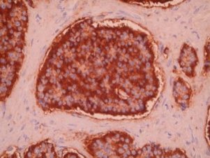
CD57 by IHC
12376 - Technical only, 12379 - Technical & interpretation
LAB12376
LAB12379
LAB12379
Leu-7
All IHC stains will include a positive control tissue
- CD57 stains the small lymphocytes in nodular lymphocyte predominance Hodgkin's disease (NLPHD), in a ring-like pattern surrounding L&H cells (this pattern can distinguish NLPHD from nodular sclerosing Hodgkin's disease, T-cell-rich B-cell lymphoma, and follicular NHL
- CD57 is useful in tumor diagnosis as a specific but relatively insensitive pan-neuroendocrine marker, usually showing positive staining in neuroblastomas, retinoblastomas, PNETs, neuroepitheliomas, carcinoids, Merkel cell tumors of the skin, paragangliomas, pheochromocytomas, small cell neuroendocrine carcinomas of the lung, urinary bladder, colon, rectum, and endometrium, medullary carcinomas of the thyroid, and some pancreatic endocrine tumors
- Occasional cases of Ewing's sarcoma, granular cell myoblastoma, Wilms' tumor, leiomyosarcoma, synovial sarcoma, epithelioid sarcoma, clear cell sarcoma, embryonal rhabdomyosarcomas, and malignant melanomas also stain positively for CD57
- Positive staining is also seen in other neural tumors such as schwannomas, neurofibromas, neurofibrosarcomas, astrocytomas and ependymomas
- A high percentage of oligodendrogliomas show positive cell surface membrane staining for CD57
- Positive staining for CD57 is seen in normal and hyperplastic prostate and prostatic adenocarcinomas
- CD57 is absent in squamous cell carcinomas and adenocarcinomas (other thanreports of rare prostate tumors as above)
Specimen
Tissue
Submit a formalin-fixed, paraffin-embedded tissue
Formalin-fixed, paraffin-embedded (FFPE) tissue block
FFPE tissue section mounted on a charged, unstained slide
Ambient (preferred)
- Unlabeled/mislabeled block
- Insufficient tissue
- Slides broken beyond repair
Performance
AHL - Immunohistochemistry
Mo - Fr
1 - 2 days
Immunohistochemical staining and microscopic examination
Clinical and Interpretive info
If requested, an interpretive report will be provided
Specifications
- Anti CD57 recognizes a 110 KD glycoprotein human lymphocyte antigen that is present on approx. 25% of adult peripheral blood mononuclear cells
- CD57 is expressed on a subset of NK cells and a subset of germinal center T-cells
- This antibody also recognizes a carbohydrate epitope on sub-populations of several cell adhesion molecules including N-CAM and myelin-associated glycoprotein (MAG)
- In normal tissues, CD57 stains a common neuro-endodermal antigen with positivity in cells of the CNS, the adrenal medulla, islets of Langerhans and the cells of the APUD system in the lung and gut
Staining pattern
- Cytoplasmic and cell surface membrane based staining
References
- Kamel OW, et al: Leu 7 (CD57) reactivity distinguishes nodular lymphocyte predominance Hodgkin's disease from nodular sclerosing Hodgkin's disease, T-cell- rich B-cell lymphoma and follicular lymphoma. American Journal of Pathology, Vol 142, 541-546, 1993.
- Arber DA, Weiss LM: CD57: a review. Appl Immuno 1995, 3:137-152.
- Michels S et al: Leu-7 in small cell neoplasms. An immunohistochemical study with ultrastructural correlations. Cancer 1987, 60:2958-2964.
Billing
88342 - 1st stain
88341 - each additional stain
88341 - each additional stain
Tracking
06/16/2017
10/18/2018
01/12/2024