CD10 by IHC-12376 - Technical only, 12379 - Technical & interpretation
Test info
CD10 by IHC
12376 - Technical only, 12379 - Technical & interpretation
LAB12376
LAB12379
LAB12379
CALLA
Common ALL antigen
Common ALL antigen
- All IHC stains will include a positive control tissue
- CD10 is expressed on precursor B-lymphoblastic lymphomas, Burkitt's lymphomas, and on some precursor T-lymphoblastic leukemia/lymphomas
- CD10 will also be expressed on many follicular lymphomas, some diffuse large B-cell lymphomas, and multiple myelomas
- CD10 expression has been seen in hairy cell leukemia and chronic myelogenous leukemia
- Endometrial stromal cells and their neoplasms (and decidua) are usually positive for CD10, which helps discriminate these neoplasms from smooth muscle tumors
- Epithelium that stains for CD10 includes breast myoepithelial cells, bile canaliculi, fibroblasts, and brush border cells of kidney and gastrointestinal tract
- Recent studies have shown that CD10 labels bile canaliculi in a similar pattern as polyclonal CEA; thus, this marker is helpful in identifying hepatocellular carcinomas from non-HCC
- CD10 is also helpful in the diagnosis of renal cell carcinoma, and some pancreatic carcinomas, since these tumors are usually positive for this marker
- Some melanomas have also been reported as positive for CD10
Specimen
Tissue
Submit a formalin-fixed, paraffin embedded tissue block
Formalin-fixed, paraffin embedded (FFPE) tissue block
FFPE tissue section mounted on a charged, unstained slide
Ambient (preferred)
- Unlabeled/mislabeled block
- Insufficient tissue
- Slides broken beyond repair
Performance
AHL - Immunohistochemistry
Mo - Fr
1 - 2 days
Immunohistochemical staining and microscopic examination
Clinical and Interpretive info
If requested, an interpretive report will be provided
Specifications
- CD10 protein (zinc metallopeptidase) is found on the surface of a wide variety of hematopoietic and non-hematopoietic tissues
- CD10 is a cell surface enzyme that reduces cellular response to peptide hormones by regulating local peptide concentration
- CD10 is expressed in early lymphoid progenitors and normal germinal center cells
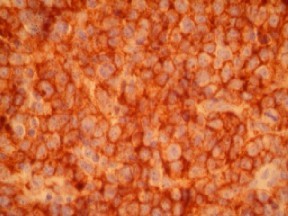
Staining pattern
- Cell surface membrane staining in lymphoid cells
- Cytoplasmic and /or membranous (including bile canalicular in HCC) in epithelial cells
References
- Kaufmann, O, et al: Immunohistochemical detection of CD10 with monoclonal antibody 56C6 on paraffin sections. Am J Clin Pathol 111; 117-122. 1999.
- Avery AK, et al: Use of antibodies to RCC and CD10 in the differential diagnosis of renal neoplasms. Am J Surg Pathol 24(2): 203-210, 2000.
- Chu PG, and Weiss LM et al: Utility of CD10 in distinguishing between endometrial stromal sarcoma and uterine smooth muscle tumors: an immunohistochemical comparison of 34 case. Mod Pathol, 14(5): 465-471, 2001.
- Notohara K, et al: Solid-pseudo-papillary tumor of the pancreas. Immunohistochemical localization of neuroendocrine markers and CD10. Am J Surg Pathol 24 (10):1361-1371, 2000.
- Borscheri N, et al: Canalicular immunostaining of Neprilysin (CD10) as a diagnostic marker for hepatocellular carcinomas. Am J Surg Pathol, 25(10): 1297-1303, 2001.
Billing
88342 - 1st stain
88341 - each additional stain
88341 - each additional stain
Tracking
05/15/2017
10/17/2018
01/12/2024