INSM1-NE by IHC-12376 - Technical only, 12379 - Technical & interpretation
Test info
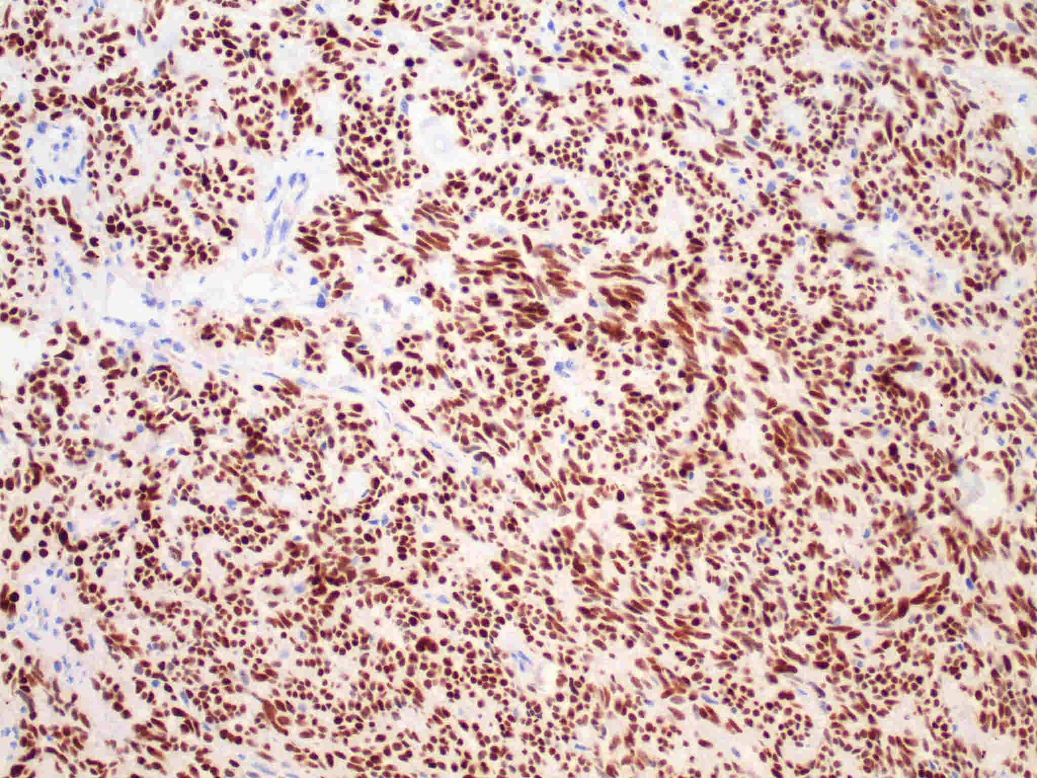
INSM1-NE by IHC
12376 - Technical only, 12379 - Technical & interpretation
LAB12376
LAB12379
LAB12379
Insulinoma-associated 1
- All IHC stains will include a positive control tissue
- INSM1 is positive in neuroendocrine tumors and in normal adult neuroendocrine tissues and developing neurons.
- INSM1 positivity has been described in the following neoplasms:
- CNS tumors: Pituitary adenoma, central neurocytoma, medulloblastoma, glioblastoma, pineal parenchymal tumor
- Endocrine: Adrenal pheochromocytoma, paraganglioma, medullary carcinoma of the thyroid, and neuroblastoma
- Gastrointestinal tract: Pancreatic neuroendocrine tumors (100%) and gastrointestinal neuroendocrine tumors (100%)
- Lung: Small cell carcinoma (95%), large cell neuroendocrine carcinoma (90%), typical and atypical carcinoid (100%). Weak or focal positivity has been reported in lung adenocarcinomas (3%) and squamous cell carcinomas (4%).
- Skin: Merkel cell carcinoma (93%), endocrine mucin producing sweat gland carcinoma
- Soft tissue tumors: Extraskeletal myxoid chondrosarcoma (90%), chordoma (10%), soft tissue myoepithelioma (5%), ossifying fibromyxoid tumor (30%), and Ewings sarcoma (30%)
- INSM1 positivity has been reported in the following in tumors with neuroendocrine differentiation: Breast adenocarcinoma, colonic adenocarcinoma, endometrioid carcinoma, and prostate adenocarcinoma
- INSM1 is positive in the following normal tissues: Adrenal medulla, enterochromaffin cells, islet cells, C cells of the thyroid, pineal gland, pituitary gland, and in neurons in early development
Specimen
Tissue
Submit a formalin-fixed, paraffin-embedded tissue
Formalin-fixed, paraffin-embedded (FFPE) tissue block
FFPE tissue section mounted on a charged, unstained slide
Ambient (preferred)
- Unlabeled/mislabeled block
- Insufficient tissue
- Slides broken beyond repair
Performance
AHL - Immunohistochemistry
Mo - Fr
1 - 2 days
Immunohistochemical staining and microscopic examination
Clinical and Interpretive info
If requested, an interpretive report will be provided
Specifications
Zinc finger transcription factor involved in development of normal neuroendocrine cells throughout the body and involved in tumor neuroendocrine differentiation.
Staining pattern
- Nuclear
References
- Ames HM et al. INSM1 expression is frequent in primary central nervous system neoplasms but not in the adult brain parenchyma. J Neuropathol Exp Neurol 2018;77(5):374-382.
- Fujino K et al. INSM1 is the best marker for the diagnosis of neuroendocrine tumors: comparison with CGA, SYP and CD56. Int J Clin Exp Pathol 2017;10(5):5393-5405.
- Lilo MT et al. INSM1 is more sensitive and interpretable than conventional immunohistochemical stains used to diagnose merkel cell carcinoma. Am J Surg Pathol 2018;42(11):1541-1548.
- Rosenbaum JN et al. A novel immunohistochemical and molecular marker for neuroendocrine and neuroepithelial neoplasms. Am J Clin Pathol 2015;144:579-591.
- Rooper LM et al. INSM1 demonstrates superior performance to individual and combined use of synaptophysin, chromogranin and CD56 for diagnosing neuroendocrine tumors of the thoracic cavity. Am J Surg Pathol 2017;41(11):1561-1569.
- Yoshinda et al. INSM1 expression and its diagnostic significance in extraskeletal myxoid chondrosarcoma. Mod Pathol 2018;31:744-752.
Billing
88342 - 1st stain
88341 - each additional stain
88341 - each additional stain
Tracking
03/04/2019
06/25/2019
01/12/2024