CD99 by IHC-12376 - Technical only, 12379 - Technical & interpretation
Test info
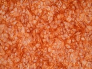
CD99 by IHC
12376 - Technical only, 12379 - Technical & interpretation
LAB12376
LAB12379
LAB12379
O13
All IHC stains will include a positive control tissue
- O13 (CD99) detects a cell surface antigen which is expressed almost exclusively on the cell membrane of Ewing's sarcoma and PNET (Primitive Neuroectodermal Tumor) but not on other small blue cell tumors (see below)
- O13 (CD99) can be useful in a panel of Immunostains for resolution of diagnostic difficulties in small blue cell tumors of childhood
- 95-100% of Ewing's sarcomas and PNETs will show positive staining for O13 (CD99)
- One must utilize O13 (CD99) in a panel as it is expressed in a wide variety of neoplasms
- Most small blue cell tumors are negative for O13 (CD99) with the exception of Acute Lymphocytic leukemia/Lymphoma. In addition, occasional rhabdomyosarcomas, Wilm's tumors and small cell osteosarcomas will show heterogeneous staining for O13 (CD99). When using a panel approach 013 can be a useful adjunct to diagnosis
- Rhabdomyosarcomas will usually express Muscle Specific Actin and desmin which Ewing's/PNET will not
- In some cases, cytogenetics may be helpful. Ewing's sarcoma and PNET usually (>90%) will show a characteristic cytogenetic abnormality t(11;22)(q24:q12)
- Reactivity of the antigen which O13 (CD99) is directed against, is maintained in decalcified tissue.2
- Up to 60% of monophasic synovial sarcomas can be O13 (CD99) positive 4, so this can be a diagnostic pitfall
Specimen
Tissue
Submit a formalin-fixed, paraffin-embedded tissue
Formalin-fixed, paraffin-embedded (FFPE) tissue block
FFPE tissue section mounted on a charged, unstained slide
Ambient (preferred)
- Unlabeled/mislabeled block
- Insufficient tissue
- Slides broken beyond repair
Performance
AHL - Immunohistochemistry
Mo - Fr
1 - 2 days
Immunohistochemical staining and microscopic examination
Clinical and Interpretive info
If requested, an interpretive report will be provided
Specifications
- O13 (CD99) detects the HBA71 antigen. HBA71 antigen is a cell surface glycoprotein (p30/32 MIC2) which is encoded by the MIC2 gene on chromosomes X and Y
- O13 (CD99) is not the only antibody which recognizes the HBA71 molecule. Others are 12E7, RFB-1 and the original antibody by the same name HBA71. These all recognize different epitopes on the same molecule. O13 (CD99) and HBA71 are most frequently used in the current literature
- Significance of O13 staining is not known
Staining pattern
- Cell membrane is typical. Cytoplasmic and perinuclear has been described
O13 (CD99) Immunoreactivity in Normal Tissue
| Strong reactivity | Variable reactivity | Focal reactivity |
| Adenohypophysis | Endothelial cells | Stomach (chief cells) |
| Ependymal cells | Fibroblasts | Bone marrow (rare lymphocytes) |
| Pancreatic islet cells | Smooth muscle | Lymph node (rare lymphocytes) |
| Granulosa cells | Skeletal muscle | Spleen (rare lymphocytes) |
| Sertoli's cells | Columnar epithelium | Kidney (collecting ducts and distal convoluted tubules) |
| Cortical thymocytes | Squamous epithelium | |
| Hassal's corpuscles | ||
| Urothelium | ||
| Myenteric plexus | ||
| Hepatocytes (fetal not adult) |
CD99 Reactivity in tumors
| 95-100% | Ewing's Sarcoma |
| 95-100% | PNET Peripheral Neuroepithelioma |
| Rare | Rhabdomyosarcoma |
| up to 90% | Lymphoblastic lymphoma |
CD99 has also been reported in:
- Alveolar Soft Parts Sarcoma
- Astrocytoma
- Embryonal carcinoma
- Ependymoma
- Glioblastoma
- Granulosa cell tumor
- Hemangiopericytoma
- Immature teratoma
- Islet cell tumor of pancreas (G+, PP+, CGA+)
- Malignant Fibrous Histiocytoma
- Melanoma
- Meningioma
- Neuroendocrine Carcinoma (carcinoids and small cell carcinoma)
- Serous Cystadenocarcinoma
- Solitary Fibrous tumor
- Squamous cell carcinoma
- Synovial Sarcoma
- Uterine Sarcoma
References
- Stevenson, AJ, Chatten J, Bertoni F and Miettinen M.; CD99 (p30/32 MIC2) Neuroectodermal/Ewing's Sarcoma Antigen as an Immunohistochemical Marker; Applied Immunohistochemistry; 2(4):231-240, 1994.
- Fellinger EJ, Garin-Chesa P, Glasser DB, Huvos AG, Rettig WJ.; Comparison of Cell Surface Antigen HBA71 (p30/32 MIC2), Neuron-Specific Enolase and Vimentin in the Immunohistochemical analysis of Ewing's Sarcoma of Bone; American Journal of Surgical Pathology; 16(*):746-755, 1992.
- Dehner LP; Primitive Neuroectodermal Tumor and Ewing's Sarcoma; American Journal of Surgical Pathology; 17(1):1-13, 1993.
- Dei Tos et. al., Immunohistochemical Demonstration of Glycoprotein P30/32MIC2(CD99) in synovial sarcoma; Applied Immunohistochemistry; 3(3):168-173, 1995.
Billing
88342 - 1st stain
88341 - each additional stain
88341 - each additional stain
Tracking
09/26/2017
10/18/2018
01/12/2022