Hepatocyte paraffin 1 by IHC-12376 - Technical only, 12379 - Technical & interpretation
Test info
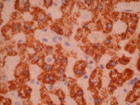
Hepatocyte paraffin 1 by IHC
12376 - Technical only, 12379 - Technical & interpretation
LAB12376
LAB12379
LAB12379
HEP PAR1
- All IHC stains will include a positive control tissue
- HepPar 1 is useful in a panel to determine hepatocellular carcinoma (HC) from cholangiocarcinoma (CC) and metastatic carcinoma
- HepPar 1 is also useful in identifying hepato-blastomas (fetal-type hepato-blastomas generally show more staining than embryonal-type)
- Some studies have shown decreased immunoreactivity with HepPar 1 in relationship to reduced differentiation of HCC
- Focal HepPar 1 immunoreactivity has also been reported in extrahepatic hepatoid adenocarcinomas, so positive staining is not limited to primary liver neoplasms(staining also has been seen in some neuroendocrine carcinomas, gastric, esophageal, lung and ovarian carcinomas)
- HepPar 1 staining may be variable in HCC; thus, needle biopsies of liver may give false negative results
- HepPar 1 can be seen in most esophageal and gastric carcinomas
Specimen
Tissue
Submit a formalin-fixed, paraffin-embedded tissue
Formalin-fixed, paraffin-embedded (FFPE) tissue block
FFPE tissue section mounted on a charged, unstained slide
Ambient (preferred)
- Unlabeled/mislabeled block
- Insufficient tissue
- Slides broken beyond repair
Performance
AHL - Immunohistochemistry
Mo - Fr
1 - 2 days
Immunohistochemical staining and microscopic examination
Clinical and Interpretive info
If requested, an interpretive report will be provided
Specifications
- HepPar 1 is an antibody that reacts with a hepatocyte-specific epitope (currently the target antigen is unknown - possibly mitochondrial associated)
- HepPar 1 reacts with both benign and malignant liver
- HepPar 1 staining rarely has been reported in cholangiocarcinomas and has been reported in mixed HCC/CC
Staining pattern
- Granular cytoplasmic
References
- Minervini MI et al: Utilization of hepatocyte-specific antibody in the immunocytochemical evaluation of liver tumors. Mod Pathol 1997;10(7):686-692.
- Kumagai I et al: Immunoreactivity to monoclonal antibody, HepPar 1 in human HCC according to the histopathological grade and histological patter. Hepatol Res 2001 Jul;20(3):312-319.
- Maitra A et al: Immunoreactivity for hepatocyte paraffin 1 antibody in hepatoid adenocarcinomas of the gastrointestinal tract. Am J Clin Pathol 2001 May;114(5): 689-94.
- Leong As et al: HepPar 1 and selected antibodies in the immunohistological distinction of MCC from CC, combined tumors and metastatic carcinoma. Histopathology 1998 Oct;33(4):318-24.
- Fasano M et al: Immunohistochemical evaluation of hepato-blastomas with use of the hepatocyte-specific marker, HepPar 1, and the polyclonal anti-CEA. Mod Pathol 1998;11(10):934-938.
- Kakar et al: Immunoreactivity of HepPar 1 in hepatic and extrahepatic tumors and its correlation with albumin in situ hybridization in HCC. Am J Clin Pathol 2003; 119: 361-366.
- Lamps LW et al: The diagnostic value of Hepatocyte Paraffin Antibody 1 in Differentiating Hepatocellular Neoplasms from Non-hepatic Tumors: A Review. Adv Anat Pathol 2003 10; 1; 39-43.
Billing
88342 - 1st stain
88341 - each additional stain
88341 - each additional stain
Tracking
07/03/2017
10/19/2018
01/12/2024