GATA3 by IHC-12376 - Technical only, 12379 - Technical & interpretation
Test info
LAB12379
All IHC stains will include a positive control tissue
Please note - GATA3 is not as specific as originally reported (see below). Also, the staining can be variable, ranging from 5% to 100%. In breast cancers, the degree of staining should parallel the ER staining. Thus, if a possible breast cancer is strongly positive for GATA3 but negative or only focally/weakly positive for ER, consider additional stains to evaluate for possible ovary or endometrial cancers.
Urothelial tumors
- GATA3 staining is found in urothelial tumors (76- 100% of urothelial cancers are GATA3 positive, per reports in literature); GATA3 is less specific but is more sensitive than uroplakin III
- GATA3 can be used to identify urothelial carcinoma, in particular when metastatic, with a higher sensitivity and specificity than either p63 or uroplakin III. This antibody is useful in a panel with PSA and PSAP to discriminate bladder from prostate cancer
- Signet ring cell adenocarcinomas / plasmacytoid carcinomas of the bladder will also stain for GATA3 (57-100%)
- GATA3 will also stain urothelial cells in Brenner tumors of the ovary, and cystic Walthard rests of the fallopian tube.
Breast tumors
- GATA3 staining is found in >90% of breast cancers; the staining is in luminal epithelium, not in myoepithelial cells
- GATA3 has comparable specificity but is more sensitive as a breast cancer marker than GCDFP-15 and mammaglobin, and can be used to discriminate lung cancers from breast cancers
Lymphoid cells
- GATA3 will stain T lymphocytes
Other tumors
- Recent articles are showing that other cancers can show more than 30% staining for GATA3, including squamous cell carcinomas and malignant mesotheliomas
- Approximately 37% of pancreatic adenocarcinomas will show positive staining for GATA3, and some renal cell carcinomas will also show positive staining (51% of chromophobe carcinomas)
- Lung adenocarcinomas can rarely show GATA3 staining (8%), as can carcinomas of the endometrium, stomach and ovary
- GATA3 staining has been reported in normal, hyperplastic and neoplastic parathyroid tissue
- Salivary gland tumors have been reported to stain for GATA3
- Some cervical and anal squamous cell carcinomas can show focal weak staining
- GATA3 has also been identified in >90% of trophoblastic and endodermal sinus tumors
- GATA3 has also been described as expressed in paragangliomas, which can be found near the bladder
- GATA3 is highly sensitive and specific for identifying mesonephric lesions in the lower genital tract.
- This marker can also be seen in basal cell carcinomas, apocrine carcinomas of the skin, and skin adnexal tumors.
Specimen
Submit a formalin-fixed, paraffin-embedded tissue
Formalin-fixed, paraffin-embedded (FFPE) tissue block
FFPE tissue section mounted on a charged, unstained slide
Ambient (preferred)
- Unlabeled/mislabeled block
- Insufficient tissue
- Slides broken beyond repair
Performance
Immunohistochemical staining and microscopic examination
Clinical and Interpretive info
If requested, an interpretive report will be provided
Specifications
- GATA-3 is a zinc finger transcription factor involved in T-cell development and in the differentiation of breast epithelium and urothelia
- Loss of GATA3 can cause a syndrome of hypoparathyroidism, sensorineural deafness, and renal dysplasia (HDR)
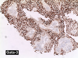
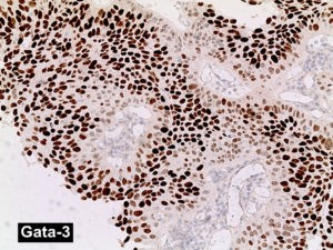
Staining pattern
- Strong nuclear staining
- Negative staining includes both negative staining and cytoplasmic (non-nuclear) staining patterns
References:
- Chang A, Amin A, Gabrielson E, Illei P, Roden RB, Sharma R, Epstein JI. Utility of GATA3 immunohistochemistry in differentiating urothelial carcinoma from prostate adenocarcinoma and squamous cell carcinomas of the uterine cervix, anus, and lung. Am J Surg Pathol. 2012 Oct;36(10):1472-6. doi: 10.1097/PAS.0b013e318260cde7. PubMed PMID: 22982890; PubMed Central PMCID: PMC3444740.
- Liu H, Shi J, Wilkerson ML, Lin F. Immunohistochemical evaluation of GATA3 expression in tumors and normal tissues: a useful immunomarker for breast and urothelial carcinomas. Am J Clin Pathol. 2012 Jul;138(1):57-64. doi: 10.1309/ AJCP5UAFMSA9ZQBZ. PubMed PMID: 22706858.
- Miyamoto H, Izumi K, Yao JL, Li Y, Yang Q, McMahon LA, Gonzalez-Roibon N, Hicks DG, Tacha D, Netto GJ. GATA binding protein 3 is down-regulated in bladder cancer yet strong expression is an independent predictor of poor prognosis in invasive tumor. Hum Pathol. 2012 Nov;43(11):2033-40. doi: 10.1016/j.humpath.2012.02.011. Epub 2012 May 18. PubMed PMID: 22607700.
- Raspollini MR, Sardi I, Giunti L, Di Lollo S, Baroni G, Stomaci N, Menghetti I, Franchi A. Plasmacytoid urothelial carcinoma of the urinary bladder: clinicopathologic, immunohistochemical, ultrastructural, and molecular analysis of a case series. Hum Pathol. 2011 Aug;42(8):1149-58. doi: 10.1016/j.humpath.2010.11.011. Epub 2011 Feb 21. PubMed PMID: 21334719.
- Raspollini MR, Comin CE, Crisci A, Chilosi M. The use of placental S100 (S100P), GATA3 and napsin A in the differential diagnosis of primary adenocarcinoma of the bladder and bladder metastasis from adenocarcinoma of the lung. Pathologica. 2010 Feb;102(1):33-5. PubMed PMID: 20731252.
- Esheba GE, Longacre TA, Atkins KA, Higgins JP. Expression of the urothelial differentiation markers GATA3 and placental S100 (S100P) in female genital tract transitional cell proliferations. Am J Surg Pathol. 2009 Mar;33(3):347-53. doi:10.1097/PAS.0b013e3181908e24. PubMed PMID: 19092634.
- Higgins JP, Kaygusuz G, Wang L, Montgomery K, Mason V, Zhu SX, Marinelli RJ, Presti JC Jr, van de Rijn M, Brooks JD. Placental S100 (S100P) and GATA3: markers for transitional epithelium and urothelial carcinoma discovered by complementary DNA microarray. Am J Surg Pathol. 2007 May;31(5):673-80. PubMed PMID: 17460449.
- Ordonez N. Value of GATA3 immunostaining in tumor diagnosis: a review. Adv Anat Pathol 2013;20:352-360.
- Miettinen M et al: GATA3: a multi-specific but potentially useful marker in surgical pathology; a systematic analysis of 2500 epithelial and nonepithelial tumors. Am J Surg Pathol 2013.
- Howitt BE et al: GATA3 Is a Sensitive and Specific Marker of Benign and Malignant Mesonephric Lesions in the Lower Female Genital Tract Am J Surg Pathol 2015;39:1411-1419).
Billing
88341 - each additional stain