P63 by IHC-12376 - Technical only, 12379 - Technical & interpretation
Test info
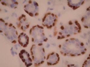
P63 by IHC
12376 - Technical only, 12379 - Technical & interpretation
LAB12376
LAB12379
LAB12379
- All IHC stains will include a positive control tissue
- P63 is most useful in breast and prostate specimens, in demonstrating the presence of an outer myoepithelial or basal layer in benign glands
- Malignant glands of prostate do not show staining with this marker; rarely, malignant breast tumor cells will be focally positive for P63
- P63 has been reported as a more sensitive marker than high molecular weight cytokeratin (HMW CK/34 beta E12) in identifying the basal layer of prostatic glands
- P63 labels most (81%) squamous cell carcinomas, and therefore can be used with CK5/6 in the identification of a poorly differentiated carcinoma in determining possible squamous epithelial origin (both markers should be positive)
- Urothelial carcinomas are often positive for P63 (70%)
- A cocktail containing both P63 and P504S (racemase) is also available in our laboratory, for the analysis of prostatic specimens
- A panel of P63 and SMMS has been recently recommend as the optimal markers for identifying the myoepithelial layer of breast<
- However, remember that P63 can demonstrate a discontinuous layer around DCIS and P63 can also rarely label breast cancer cells
- P63 is useful in distinguishing acinic cell carcinoma and metastatic renal cell carcinoma from other salivary gland tumors with clear cell and oncocytic features. It shows patchy staining in many salivary gland tumors including mucoepidermoid carcinomas, pleomorphic adenomas, basal cell adenomas, adenoid cystic carcinomas, oncocytomas, and epithelial-myoepithelial carcinomas. It is negative in acinic cell carcinomas and in metastatic renal cell carcinoma
Specimen
Tissue
Submit a formalin-fixed, paraffin embedded tissue block
Formalin-fixed, paraffin embedded (FFPE) tissue block
FFPE tissue section mounted on a charged, unstained slide
Ambient (preferred)
- Unlabeled/mislabeled block
- Insufficient tissue
- Slides broken beyond repair
Performance
AHL - Immunohistochemistry
Mo - Fr
1 - 2 days
Immunohistochemical staining and microscopic examination
Clinical and Interpretive info
If requested, an interpretive report will be provided
Specifications
- P63 is a member of the P53 family, is expressed in the basal cells of many types of epithelia, including skin, cervix, prostate, and GU tract
Staining pattern
- Nuclear staining
References
- Barbareschi M. et al: P63, a P53 homologue, is a selective nuclear marker of myoepithelial cells of the human breast. Am J Surg Pathol 25(8): 1054-1060, 2001.
- Kaufmann O. et al: Value of P63 and cytokeratin 5/6 as immunohistochemical markers for the differential diagnosis of poorly differentiated and undifferentiated carcinomas. Am J Clin Pathol 2001;116; 823-830.
- Shah RB et al: Comparison of the basal cell-specific markers, 34BE12 and P63, in the diagnosis of prostate cancer. Am J Surg Pathol 26(9): 1161-1168, 2002.
- Wang BY et al: P63 in pulmonary epithelium, pulmonary squamous neoplasms, and other pulmonary tumors. Human Pathol, 33(9), 921-926, 2002.
- Werling RW et al: Immunohistochemical distinction of invasive from noninvasive breast lesions; a comparative study of )63 vs. calponin and smooth muscle myosin heavy chain. Am J surg Pathol 27(1): 82-90, 2003.
- Bilal H, Handra-Luca A, Bertrand JC, Fouret PJ P63 is expressed in basal and myoepithelial cells of human normal and tumor salivary gland tissues J Histochem Cytochem. 2003 Feb;51(2):133-9.
- Sams RN, Gnepp DR P63 expression can be used in differential diagnosis of salivary gland acinic cell and mucoepidermoid carcinomas. Head Neck Pathol. 2013 Mar;7(1):64-8.
- Carvalho JC, Thomas DG, McHugh JB, Shah RB, Kunju LP p63, CK7, PAX8 and INI-1: an optimal immunohistochemical panel to distinguish poorly differentiated urothelial cell carcinoma from high-grade tumours of the renal collecting system. Histopathology. 2012 Mar;60(4):597-608.
Billing
88342 - 1st stain
88341 - each additional stain
88341 - each additional stain
Tracking
07/19/2017
10/19/2018
01/12/2024