CD33 by IHC-12376 - Technical only, 12379 - Technical & interpretation
Test info
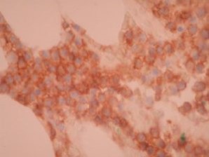
CD33 by IHC
12376 - Technical only, 12379 - Technical & interpretation
LAB12376
LAB12379
LAB12379
All IHC stains will include a positive control tissue
- CD33 is useful in the identification of cells of myeloid and monocytic lineage, and of leukemias and myeloproliferative disorders derived from these cells, including acute myeloid leukemias, chronic myeloid leukemias, granulocytic sarcomas, myeloid dysplasia and myeloproliferative diseases
- CD33 staining may rarely be seen in precursor B-lymphoblastic leukemias, precursor T-lymphoblastic leukemias, and rare cases of classical Hodgkins disease, Burkitt lymphoma, plasma cell neoplasms
- Identifying CD33 expression is necessary for targeted therapy using anti-CD33 drugs (gemtuzumab ozogamicin/Mylotarg)
- This antibody may be useful in minimally differentiated AML-M0 or AML-M5 where myeloperoxidase may be negative
Specimen
Tissue
Submit a formalin-fixed, paraffin-embedded tissue
Formalin-fixed, paraffin-embedded (FFPE) tissue block
FFPE tissue section mounted on a charged, unstained slide
Ambient (preferred)
- Unlabeled/mislabeled block
- Insufficient tissue
- Slides broken beyond repair
Performance
AHL - Immunohistochemistry
Mo - Fr
1 - 2 days
Immunohistochemical staining and microscopic examination
Clinical and Interpretive info
If requested, an interpretive report will be provided
Specifications
- CD33 is a protein found in the C2 domain of human CD33, mapped to chromosome 19q13.1-3
- This antigen may possibly have a role in cell-to-cell adhesion, and is present only on committed myeloid precursor stem cells
- CD33 is present in cells of myelomonocytic lineages, and also stains mature macrophages; cells of erythroid and megakaryocytic lineage are negative for this protein
- Staining has been reported in placental syncytiotrophoblasts; no other non-hematopoietic staining has been reported in other epithelial or mesenchymal tissues
Staining pattern
- Cell surface membrane and cytoplasmic
References
- Hoyer JD et al: CD33 detection by immunohistochemistry in paraffin-embedded tissues: a new antibody shows excellent specificity and sensitivity for cells of myelomonocytic lineage.Am J Clin Pathol. 2008 Feb;129(2):316-23.
- Yeung J et al: Genomic aberrations and immunohistochemical markers as prognostic indicators in multiple myeloma. J Clin Pathol. 2007 Dec 21.
Billing
88342 - 1st stain
88341 - each additional stain
88341 - each additional stain
Tracking
06/16/2017
10/17/2018
01/12/2024