IgA by IHC-12376 - Technical only, 12379 - Technical & interpretation
Test info
IgA by IHC
12376 - Technical only, 12379 - Technical & interpretation
LAB12376
LAB12379
LAB12379
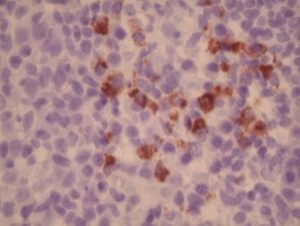
Immunoglobulin A
- All IHC stains will include a positive control tissue
- IgA labels plasma cells and related lymphoid cells containing IgA
- IgA stains will show an interstitial dendritic pattern within lymphoid follicles, (this represents extracellular immune complexes which are bound to the surface of specialized follicular dendritic cells)
- IgA will label occasional B cell lymphomas
Specimen
Tissue
Submit a formalin-fixed, paraffin-embedded tissue
Formalin-fixed, paraffin-embedded (FFPE) tissue block
FFPE tissue section mounted on a charged, unstained slide
Ambient (preferred)
- Unlabeled/mislabeled block
- Insufficient tissue
- Slides broken beyond repair
Performance
AHL - Immunohistochemistry
Mo - Fr
1 - 2 days
Immunohistochemical staining and microscopic examination
Clinical and Interpretive info
If requested, an interpretive report will be provided
Specifications
- This antibody reacts with heavy chains (alpha) of human IgA (Fc-part)
- This antibody labels plasma cells and various lymphocytes
Staining pattern
- Cytoplasmic (plasma cells) and cell surface membrane (lymphocytes)
References
- Taylor CR et al: The demonstration of plasma cells and other immunoglobulin-containing cells in formalin-fixed, paraffin-embedded tissues using peroxidase-labelled antibody. J Clin Pathol 1974; 27:14-20.
- Taylor CR et al: The immunohistological detection of intracellular immunoglobulin in formalin-paraffin sections from multiple myeloma and related conditions using the immunoperoxidase technique. Clin exp Immunol 1974; 8:417-29.
Billing
88342 - 1st stain
88341 - each additional stain
88341 - each additional stain
Tracking
07/03/2017
10/19/2018
01/12/2024