Granzyme B by IHC-12376 - Technical only, 12379 - Technical & Interpretation
Test info
Granzyme B by IHC
12376 - Technical only, 12379 - Technical & Interpretation
LAB12376
LAB12379
LAB12379
All IHC stains will include a positive control tissue
- Granzyme B is used (often in a panel with T1A-1 and/or perforin) to identify cytotoxic (NK-like) T-cell lymphomas, especially enteropathy associated T-cell lymphomas, and subcutaneous panniculitis-like T-cell lymphoma. These antibodies also identify true NK cell lymphomas/leukemias, including aggressive NK-cell leukemia/lymphoma and extra-nodal NK/T-cell lymphoma, nasal type
- NK cell lymphomas are usually aggressive, and are associated with rapid progression, treatment failures, and relapses; therefore, accurate diagnosis is essential
- Some studies report an associated of nasal-type NK cell lymphomas with EBV, and are more common in Asian and Latin American countries
- P53 is also expressed in a high percentage of these types of lymphomas
- Granzyme B should be used in a panel with other T cell markers, as well as with CD56 and T1A-1 and or perforin
Specimen
Tissue
Submit a formalin-fixed, paraffin-embedded tissue
Formalin-fixed, paraffin-embedded (FFPE) tissue block
Tissue section mounted on a charged, unstained slide
Ambient (preferred)
- Unlabeled/mislabeled block
- Insufficient tissue
- Slides broken beyond repair
Performance
AHL - Immunohistochemistry
Mo - Fr
1 - 2 days
Immunohistochemical staining and microscopic examination
Clinical and Interpretive info
If requested, an interpretive report will be provided
Specifications
- Granzyme B is a granule-associated serine protease type of protein, found in cytotoxic T lymphocytes (CTL) and in natural killer (NK) cells
- Granzyme B and perforin are essential for apoptosis and immune-mediated cell death of target cells via induction of cell membrane perforation
- Granzyme B expression is evidence of an activated state of CTL and NK cells, and is therefore useful in identifying related lymphomas associated with these types of cells
- Expression of this marker has been associated with a poor prognosis in nodular sclerosis or mixed cellularity Hodgkin's disease
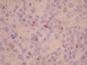
Staining pattern
- Granular cytoplasmic staining
References
- Gall K et al: Sinonasal NK/T-cell lymphomas in the United States. Am J Surg Pathol 24(11):1511-1517, 2000.
- Natkunam Y et al: Aggressive cutaneous NK and NK-like T-cell lymphomas; clinicopathologic, immunohistochemical and molecular analyses of 12 cases. Am J Surg Pathol 23(5): 571-581, 1999.
- Quintanilla-Martinez L et al: Histological and Immunophenotypic profile of nasal NK/T cell lymphomas from Peru: high prevalence of p53 over-expression. Hum Pathol 30:849-855, 1999.
- Kumar S et al: Subcutaneous panniculitic T-cell lymphoma is a tumor of cytotoxic T lymphocytes. Hum Pathol 29:397-403, 1998.
Billing
88342 - 1st stain
88341 - each additional stain
88341 - each additional stain
Tracking
07/03/2017
10/18/2018
01/12/2024