BCL-1 by IHC-12376 - Technical only, 12379 - Technical & interpretation
Test info
LAB12379
- All IHC stains will include a positive control tissue
- Cyclin D1/PRAD-1 plays a role in the regulation of the eukaryotic cell cycle, maximum expression occurring in the mid to late GI phase of the cycle. Over expression of Cyclin D1 protein is thought to contribute to tumorigenesis by deregulating the normal cell cycle. The Cyclin D1 gene is located on 11q13
- In lymphomas, Cyclin D1 over expression usually occurs as a result of the bcl-1 rearrangement t(11;14)(q13;q32)
- Normal or reactive lymphoid tissue and hematopoietic cells do not show Cyclin D1 over expression (only very few cells localized in the mantle zones express Cyclin D1).
- bcl-1 reactivity is seen in nearly 90-100% of cases of mantle cell lymphoma. (The t(11;14) is seen in approximately 70% of MCL). Over expression of PRAD-1/Cyclin D1 more essential to defining MCL than CD5
- The t(11;14) or bcl-1 rearrangement is rarely found in other lymphoid malignancies and the frequency of Cyclin D1 over expression in other lymphomas supports that observation
Table 1 (Ref 4) Cyclin D1 expression in lymphomas:
| Low grade B-cell neoplasms | 6/38 |
| B-chronic lymphocytic leukemia/ small lymphocytic lymphoma |
0/18 |
| Lymph node | 0/13 |
| Soft tissue | 0/3 |
| Spleen | 0/1 |
| Stomach | 0/1 |
| Mantle cell lymphoma | 5/5 |
| Lymph node | 4/4 |
| Colon (lymphomatous polyposis) | 1/1 |
| Hairy cell leukemia (spleen) | 1/15 |
| Plasmacytoma/plasma cell myeloma (19 bone, 3 head and neck) |
1/22 |
| High grade lymphomas | 0/46 |
| Diffuse large B-cell lymphoma | 0/33 |
| Lymph node | 0/11* |
| Bone | 0/9 |
| Tonsil/nasopharynx | 0/6* |
| Mediastinum | 0/1 |
| Mesentery/pericolonic | 0/2 |
| Lung | 0/2 |
| Soft tissue | 0/2 |
| Burkitt's and Burkitt-like lymphomas | 0/9 |
| Lymph node | 0/3 |
| Mesentery/omentum | 0/3 |
| Nasopharynx/salivary gland | 0/2 |
| Soft tissue | 0/1 |
| Precursor T-Lymphoblastic lymphoma | 0/4 |
| Lymph node | 0/4 |
| Adult T-cell lymphoma/leukemia Lymphomatous type (bone) |
0/1 |
*CD5+ B-cell lymphomas involved lymph nodes (2 cases) and tonsil (1 case).
Table 2 (Ref 1) Results of PRAD1/CYCLIN D1 staining:
Cyclin D1-positive (nuclear positive pattern) Non-Hodgkin’s lymphomas (total 238 cases)
B-lineage Follicular centroblastic-centrocytic
| lymphoma | 0/34 (0%)a |
| Small lymphocytic lymphoma, CLL | 0/8 (0%) |
| Mantle cell lymphoma (centrocytic) | 35/39(90%)b |
| Low grade B-cell lymphoma with MCL morphology and absence of CD5 expression | 0/4 (0%)c |
| Immunocytoma | 1/6 (17%)d |
| Plasmacytic lymphoma (plasmacytoma) | 0/4 (0%) |
| Marginal zone/monocytoid B-cell lymphoma | 0/10 (0%) |
| Nodal, 6 cases | 0/6 |
| Extranodal, 3 tonsil, 1 spleen | 0/4 |
| Low-grade B-cell lymphoma of MALT type | 0/39 (0%) |
| Diffuse centroblastic/immunoblastic lymphoma | 0/90 (0%) |
| Nodal | 0/52e |
| Extranodal (30 GI tract, 2 breast, etc) | 0/38 |
| Burkitt’s lymphoma, extranodal (2 stomach, 1 tonsil) | 0/3 (0%) |
| T/NK-lineage | |
| T-lymphoblastic lymphoma | 0/6 (0%) |
| Other T/NK-cell lymphomas | 0/34 (0%) |
| Hodgkin’s disease (total 19 cases) | 1/19 (5%)f |
| Reactive lesions | 0/37 (0%) |
| TOTAL | 334 cases |
CLL, chronic lymphocytic leukemia; MCL, mantle cell lymphoma; MALT, mucosa-associated lymphoid tissue; GI, gastrointestinal; NK, natural killer.
a - cases positive/cases examined.
b - consisted of 35 Group I cases and four Group II cases (see Fig. 1).
c - corresponded to four Group III cases.
d - one Cyclin D1-positive case was included in Group I.
e - included 11 cases of CD5-positive tumors defined as Group IV.
f - a few mummified Reed-Sternberg cells showed nuclear staining.
- Cyclin D1 over expression is also seen in parathyroid adenomas (associated with the chromosomal translocation), breast (>33%) and squamous cell carcinoma of head and neck (associated with amplification of the 11q13 region)
- In some breast carcinomas, Cyclin D1 may be over expressed in the absence of translocation (with or without 11q13 amplification) raising the possibility that some lymphomas lacking the translocation may express the protein
- Cross reactivity may be seen with endothelial cell nuclei and cytoplasm and fibroblasts in some cases
Specimen
Submit a formalin-fixed, paraffin embedded tissue block
Formalin-fixed, paraffin embedded (FFPE) tissue block
Tissue section mounted on a charged, unstained slide
Ambient (preferred)
- Unlabeled/mislabeled block
- Insufficient tissue
- Slides broken beyond repair
Performance
Immunohistochemical staining and microscopic examination
Clinical and Interpretive info
If requested, an interpretive report will be provided
Specifications
Reacts with human Cyclin D1 in formalin fixed or frozen tissue. The DCS6 antibody does not cross react with Cyclin D2 and/or Cyclin D2, both of which are highly expressed in many normal tissues (including lymphoid tissue) and tumors.
Staining pattern
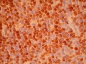
- Granular to diffuse nuclear staining (seen in 20-90% of lymphoid cells)
- Pronounced variation in staining intensity observed among individual tumor cells
References
- AJSP 20 (9):1110-1122, 1996.
- AJSP 20 (5):627-640, 1996.
- Blood Vol86 (7):2715-2723, Oct., 1995.
- AJCP 103:756-760, 1995.
- Human Pathology, 26:999-1004.
- Applied Immunohistochemistry, 5(1):45-48, 1997.
- Novocastra and Oncogene spec sheets.
Billing
88341 - each additional stain