BCL-2 by IHC-12376 - Technical only, 12379 - Technical & interpretation
Test info
BCL-2 by IHC
12376 - Technical only, 12379 - Technical & interpretation
LAB12376
LAB12379
LAB12379
- All IHC stains will include a positive control tissue
- Bcl-2 protein is the protein product of the oncogene trans-located in most follicular lymphomas t(14;18). It is an inner mitochondrial membrane protein that blocks programmed cell depth and therefore, cells that over express this protein have a survival advantage over other cells
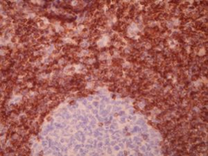
- In normal lymph nodes, positive staining is seen in the mantle zone cells, many interfollicular T and B cells, and in scattered follicular cells
- In bone marrow, Bcl-2 also stains plasma cells, mast cells, granulocytes, and dendritic cells
- In neoplastic lymph nodes, Bcl-2 expression has been seen in lymphomas containing the t(14;18) translocation as well as in most follicular lymphomas. One study reported 100% of follicular small cleaved lymphomas staining for Bcl-2, and a lower proportion of follicular mixed, follicular large cell and large cell non-cleaved lymphomas (86% follicular mixed cell type; 76% follicular large cell type). Bcl-2 also labels hairy cell leukemia, some high grade B and T cell non-Hodgkin’s lymphomas, lymphoblastic lymphomas and Ki-1 lymphomas. However, Bcl-2 expression is not specific for t(14;18) and is seen in normal B and T cells as well as in follicular lymphomas and non-follicular lymphomas which do not have the t(14;18)
- Bcl-2 reactivity would be most helpful in determining reactive follicular hyperplasia from follicular lymphoma. Because Bcl-2 reactive T-cells are present in germinal centers, there is a need to control for over interpretation of Bcl-2 by doing a CD3. Then, follicles judged to be Bcl-2 positive in proportions greater than the CD3 positive cells are favored for neoplastic1,2,3
- In bone marrow, this antibody may help distinguish between benign and malignant lymphoid aggregates. One study reports that the lack of Bcl-2 staining in the lymphoid aggregate favors a benign lymphoid population. Moderate to strong staining was reported to favor a malignant lymphoid aggregate 4
References:
- Utz GL and Swerdlow SH; Distinction of Follicular Hyperplasia from Follicular Lymphoma in B5-Fixed Tissues: Comparison of MT2 and Bcl-2 Antibodies; Human Pathology 24(11):1155-1158, 1993.
- Banks PM; When is a New Diagnostic Method Established?; Human Pathology 24(11):1153-1154, 1993.
- Wood BL, Bacchi MM, et. al.; Immunocytochemical Differentiation of Reactive Hyperplasia from Follicular Lymphoma Using Monoclonal Antibodies to Cell Surface and Proliferation-Related Markers; Applied Immunohistochemistry 2(1):48-53, 1994.
- Ben-Ezra JM, King BE, et. al.; Staining for Bcl-2 Protein Helps to Distinguish Benign from Malignant Lymphoid Aggregates in Bone Marrow Biopsies; Modern Pathology 7(5):560-564, 1994.
Specimen
Tissue
Submit a formalin-fixed, paraffin embedded tissue block
Formalin-fixed, paraffin embedded (FFPE) tissue block
Tissue section mounted on a charged, unstained slide
Ambient (preferred)
- Unlabeled/mislabeled block
- Insufficient tissue
- Slides broken beyond repair
Performance
AHL - Immunohistochemistry
Mo - Fr
1 - 2 days
Immunohistochemical staining and microscopic examination
Clinical and Interpretive info
If requested, an interpretive report will be provided
Specifications
- Reacts with Bcl-2 oncoprotein in formalin fixed or in B3/B5 fixed paraffin embedded tissue
Staining patterns
- Cell surface membrane and cytoplasmic staining
Billing
88342 - 1st stain
88341 - each additional stain
88341 - each additional stain
Tracking
05/10/2017
10/17/2018
01/12/2024