PAX8 by IHC-12376 - Technical only, 12379 - Technical & interpretation
Test info
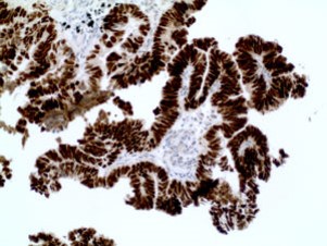
PAX8 by IHC
12376 - Technical only, 12379 - Technical & interpretation
LAB12376
LAB12379
LAB12379
- All IHC stains will include a positive control tissue
- PAX8 is expressed in ovarian serous (99%), endometrioid (98%), and clear cell carcinomas (78%) (and only very rarely in mucinous ovarian tumors), and cervical carcinomas (91%)
- PAX8 is present in 90-98% of clear cell renal cell carcinomas, 90% of papillary renal cell carcinomas, and 81-95 % of oncocytomas
- PAX8 is expressed in thyroid neoplasms (91%)
- No (or focal weak) expression for PAX8 is seen in lung, breast, and non-GYN carcinomas
- Thymic carcinomas and thymomas may show moderate staining with PAX8
- PAX8 is useful in a panel differentiating peritoneal mesothelioma from papillary serous
- PAX8 can be used to discriminate GYN tumors from breast tumors
Specimen
Tissue
Submit a formalin-fixed, paraffin embedded tissue block
Formalin-fixed, paraffin embedded (FFPE) tissue block
FFPE tissue section mounted on a charged, unstained slide
Ambient (preferred)
- Unlabeled/mislabeled block
- Insufficient tissue
- Slides broken beyond repair
Performance
AHL - Immunohistochemistry
Mo - Fr
1 - 2 days
Immunohistochemical staining and microscopic examination
Clinical and Interpretive info
If requested, an interpretive report will be provided
Specifications
- PAX8 is a member of the PAX family of transcription factors that are active in thyroid development
- PAX8 expression is seen in thyroid, non-ciliated mucosal cells of fallopian tubes, ovarian inclusion cysts, and in renal tubular cells and associated tumors
- PAX8 positivity can be seen in B-cell lymphomas, likely due to cross reactivity with PAX5
Staining pattern
- Nuclear
References
- Laury AR et al: A Comprehensive Analysis of PAX8 Expression in Human Epithelial Tumors Am J Surg Pathol 2011;35:816-826).
- Morgan EA at al: PAX8 and PAX5 are differentially expressed in B-cell and T-cell lymphomas. Histopathology 2013, 62, 406-413.
- Moretti L at al: N-terminal PAX8 polyclonal antibody shows cross-reactivity with N-terminal region of PAX5 and is responsible for reports of PAX8 positivity in malignant lymphomas. Modern Pathology 2012, 25, 231-236.
- Ordonez NG: Value of PAX 8 Immunostaining in Tumor Diagnosis: A Review and Update. Adv Anat Pathol 2012;19:140-151).
Billing
88342 - 1st stain
88341 - each additional stain
88341 - each additional stain
Tracking
08/07/2017
10/19/2018
01/12/2024