Alpha-1-antitrypsin by IHC-12376 - Technical only, 12379 - Technical & interpretation
Test info
Alpha-1-antitrypsin by IHC
12376 - Technical only, 12379 - Technical & interpretation
LAB12376
LAB12379
LAB12379
AAT
IHC
IHC
- All IHC stains will include a positive control tissue
- The primary use for this antibody is in identifying the accumulation of AAT granules in alpha-1-antitrypsin deficiency of the liver
- AAT is a very useful marker in the work-up of alpha-1-antitrypsin deficiency, since although it is synthesized by the liver in this disease, it is not released into the circulation (thus, the serum is deficient)
- This antibody is also useful in identifying tumors which can make this protein, such as liver neoplasms, endodermal sinus tumors, and for histiocytic related tumors
- AAT is expressed in liver related proliferations as follows: focal nodular hyperplasia (56%), liver cell adenoma (68%) and hepatoma (89%)
Specimen
Tissue
Submit a formalin-fixed, paraffin embedded tissue block
Formalin-fixed, paraffin embedded (FFPE) tisue block
Tissue section mounted on a charged, unstained slide
Ambient (preferred)
- Unlabeled/mislabeled
- Insufficient tissue
- Slides broken beyond repair
Performance
AHL - Immunohistochemistry
Mo - Fr
1 - 2 days
Immunohistochemical staining and microscopic examination
Clinical and Interpretive info
If requested, an interpretive report will be provided
Specifications
- AAT is a glycoprotein that represents a major component of the alpha-1-globulin serum fraction (90%)
- AAT is found in serum, as well as in other bodily fluids such as lymphatics, mucus, saliva, synovial fluid, GI secretions, semen, amniotic fluid and colostrum
- The embryonal yolk sac is the initial site of AAT production
- Although AAT is predominately made in the liver, it is also produced in histiocytes and monocytes
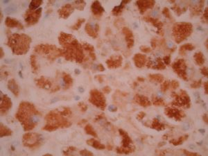
Staining patterns
- Granular based cytoplasmic activity
- In alpha-1-antitrypsin deficiency, the staining appears as large globules, particularly around the periphery of hepatocytes
References
- Palmer, P.E. et al; Am J Surg Pathol 2:275-281, 1978.
Billing
88342 - 1st stain
88341 - each additional stain
88341 - each additional stain
Tracking
03/02/2016
01/31/2024
01/31/2022