Epithelial membrane antigen by IHC-12376 - Technical only, 12379 - Technical & interpretation
Test info
Epithelial membrane antigen by IHC
12376 - Technical only, 12379 - Technical & interpretation
LAB12376
LAB12379
LAB12379
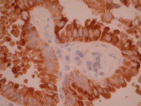
EMA
All IHC stains will include a positive control tissue
- Breast and skin adnexa are usually strongly EMA positive
- Variety of other carcinomas that are EMA positive include carcinomas of endometrium, kidney, thyroid, stomach, pancreas, lung, colon, ovary, prostate, cervix
- EMA positivity may be seen in mesotheliomas; transitional and squamous cell carcinomas may show variable reactivity
- EMA also labels plasma cells, NHL with plasmacytic differentiation, and anaplastic large cells lymphomas (Ki-1 lymphomas)
- Hepatocellular carcinomas, adrenocortical carcinomas, embryonal carcinomas and basal cell carcinomas are (usually) negative for EMA
EMA positivity in NHL and HD:
| B cell NHL | 5% |
| Plasma cell | 80% |
| T cell NHL | 20% |
| Anaplastic (Ki-1) | 80% |
| L & H nodular HD | 60% |
| Classical HD | 20% |
Specimen
Tissue
Submit a formalin-fixed, paraffin-embedded tissue
Formalin-fixed, paraffin-embedded (FFPE) tissue block
FFPE tissue section mounted on a charged, unstained slide
Ambient (preferred)
- Unlabeled/mislabeled block
- Insufficient tissue
- Slides broken beyond repair
Performance
AHL - Immunohistochemistry
Mo - Fr
1 - 2 days
Immunohistochemical staining and microscopic examination
Clinical and Interpretive info
If requested, an interpretive report will be provided
Specifications
- EMA is a glycoprotein isolated from human milk fat globule membranes
- EMA is present in many types of epithelia
- EMA can also be identified in mesenchymal and hematopoietic cells
Staining pattern
- Cytoplasmic based staining and +/- cell surface membrane staining
Billing
88342 - 1st stain
88341 - each additional stain
88341 - each additional stain
Tracking
06/21/2017
10/17/2018
01/12/2024