CD56 by IHC-12376 - Technical only, 12379 - Technical & interpretation
Test info
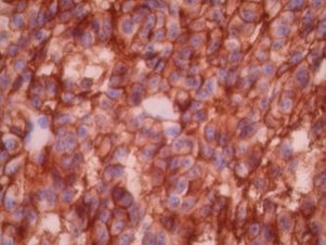
CD56 by IHC
12376 - Technical only, 12379 - Technical & interpretation
LAB12376
LAB12379
LAB12379
NCAM
Neural cell adhesion molecule
Neural cell adhesion molecule
All IHC stains will include a positive control tissue
- For the diagnosis of nasal and extra-nasal CD-56 positive lymphomas (these have a characteristic clinical picture, sites of involvement, immunophenotype CD2+, CD3-, CD56+, negative TCR, and Ig gene rearrangements, worse prognosis and a very aggressive behavior. Also, they are ·frequently EBV positive) 4, 5
- Ab can be used to support a diagnosis or to detect minimal residual disease in these cases of CD-56 and NK cell lymphoma
- CD-56 also positive in some other T-cell leukemias/lymphomas. e.g.:
- Hepatosplenic gamma-delta T-cell lymphoma (usually CD4-, CD8-, CD3+, and consistently show TCR-delta gene rearrangements)
- S-100 and T-cell lymphoproliferative disorder (CD4-, CD8+, S-100+, CD16+, CD56+, TCR? /?, GR+, EBV-)
- In general B cell lymphomas lack CD56 expressions. 1
- CD-56 is the most commonly expressed among the available natural killer cell markers
- <0.1% of all lymphocytes in normal or reactive lymph nodes, nasal/nasopharyngeal mucosa, tonsils, thymus, and appendix show positive staining. These positive cells occur singly or at most in tiny groups of 2-3 cells (unlike neoplastic cells)
- Small fraction of plasma cells (1-5%) show weak to moderate cytoplasmic staining
- CD-56 expression is seen in other hemato-lymphoid malignancies and is frequently associated with a worse prognosis (e.g. ALL, AML, multiple myeloma, CD3+, T-cell LGL and lymphoblastic lymphoma) 1, 5
- Small cell carcinomas and carcinoids of the lung are strongly positive in all cells
- Some rhabdomyosarcomas, malignant melanomas and Wilms tumors are also positive2
- Several cases of adenoid cystic carcinomas of bronchial glands are strongly positive
- Some medullary thyroid carcinomas and ovarian tumors are positive8
- Nerve fibers are strongly positive and serve as good internal controls
- Other cell types, including sub-populations of breast, prostate, testicular epithelium and ovarian and prostatic interstitial cells have also been found to react in formalin fixed tissue7,8
Specimen
Tissue
Submit a formalin-fixed, paraffin-embedded tissue
Formalin-fixed, paraffin-embedded (FFPE) tissue block
FFPE tissue section mounted on a charged, unstained slide
Ambient (preferred)
- Unlabeled/mislabeled block
- Insufficient tissue
- Slides broken beyond repair
Performance
AHL - Immunohistochemistry
Mo - Fr
1 - 2 days
Immunohistochemical staining and microscopic examination
Clinical and Interpretive info
If requested, an interpretive report will be provided
Specifications
- The antibody recognizes the 14-KD isoform of the neural cell adhesion molecule (NCAM) which is a surface glycoprotein found in natural killer (NK) cells, NK like T-cells, muscle and neural/neuroendocrine tissues and their corresponding neoplasms (e.g. pheochromocytoma, neuroblastoma, medulloblastoma, astrocytoma, retinoblastoma and small cell carcinoma).
Staining pattern
- Cell membrane
- With the clone 123C3 true positive cells form multiple sheets and compact clusters comprising more than 6 cells
- >20% of the neoplastic lymphoid cells should stain with the Ab1
References
- AJSP 20(2):202-210, 1996.
- Leukemia and Lymphoma, 14:29-3, 1993.
- Blood; 85(6):1675-6, 1995.
- AJSP; 19(3):284-296, 1995.
- Human Pathology; 23:798-804, 1992.
- Leukemia and lymphoma; 19:295-300, 1995.
- DAKO specification sheet.
- Accurate Chem specification sheet.
Billing
88342 - 1st stain
88341 - each additional stain
88341 - each additional stain
Tracking
06/16/2017
10/18/2018
01/12/2024