Protein S, free-5750
Test info
Protein S, activity
Warfarin (Coumadin)
Specimen
Warfarin (Coumadin) therapy should be discontinued two weeks prior and heparin therapy two days prior to sample collection.
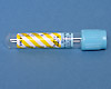
Lt blue Sodium citrate (Na Cit) - 2.7mL
If the patient has a hematocrit >55, a specially prepared Lt blue Sodium citrate (NaCit) tube must be used in place of the standard Lt blue Sodium citrate (NaCit) tube.
- Do not draw from an arm with a heparin lock or heparinized catheter
- Do not over or under fill tube as the ratio of anticoagulant to whole blood is critical
- Immediately following collection, mix sample thoroughly by gently inverting 8 - 10 times to prevent clotting
- Process Platelet Poor Plasma (P.P.P)
- Transfer plasma into an aliquot tube using a spot label "NaCit Plasma P.P.P" to label as platelet poor plasma
- Freeze immediately
Microcentrifuge vial/tube in a Snap cap conical vial/tube and cap (Beaker sites)
Coagulation specimen transport vial/tube (all other sites)
Frozen (strict)
Refrigerated - NO
- Greiner tubes are not acceptable
- Improper labeling (unlabeled or mislabeled)
- Improper anticoagulant
- Improperly filled tube
- Hemolysis
- Clotted specimen
- Delay in transport
- Improper storage/transport temperature
- Patient on heparin > 1.0 IU/mL
Performance
Available STAT
Protein S, free – Immuno-turbidometric
Clinical and Interpretive info
Protein S, free (Antigenic):
Female: 55-124%
Male: 74-126%
Guidance for the evaluation and treatment of hereditary and acquired thrombophilia
|
Types of Heterozygous Protein S deficiency |
|||
|
|
Protein S, free Ag |
Protein S, total |
Protein S, activity |
|
Type I |
Decreased |
Decreased |
Decreased |
|
Type II |
Normal |
Normal |
Decreased |
|
Type III |
Decreased |
Normal |
Decreased |
The three subtypes of heritable protein S deficiencies include both qualitative and quantitative defects. Type I is a quantitative defect resulting in decreased plasma antigen levels of both free and total protein S antigen. Type II is a qualitative defect with decreased protein S activity and normal levels of free and total protein S antigen. Type III is characterized by decreased free protein S antigen and protein S activity with normal levels of total protein S antigen. Type I and III protein S deficiency are much more common than type II (dysfunctional) protein S deficiency.
Acquired free protein S deficiency may be seen in certain inflammatory conditions and will most often have decreased levels of free protein S antigen and protein S activity, but normal to elevated total protein S antigen. The hemostatic implications for patients with acquired protein S deficiency are of unclear clinical significance.
Reference:
Quality in Laboratory Hemostasis and Thrombosis. Kitchen, Steve, Olson, John D., and Preston, F. Eric. 2009 Blackwell Publishing Ltd, Sussex, UK. pg. 152.