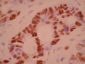
p53 by IHC
p53 by IHC-12376 - Technical only, 12379 - Technical & interpretation
p53 by IHC
12376 - Technical only, 12379 - Technical & interpretation
LAB12376
LAB12379
LAB12379
- All IHC stains will include a positive control tissue
P53 over-expression is present in many different malignancies, such as colon, stomach, bladder, breast, lung, testes, melanomas, sarcomas, etc.
Tissue
Submit a formalin-fixed, paraffin embedded tissue block
Formalin-fixed, paraffin embedded (FFPE) tissue block
FFPE tissue section mounted on a charged, unstained slide
Ambient (preferred)
- Unlabeled/mislabeled block
- Insufficient tissue
- Slides broken beyond repair
AHL - Immunohistochemistry
Mo - Fr
1 - 2 days
Immunohistochemical staining and microscopic examination
If requested, an interpretive report will be provided
Specifications
- P53 is a transcription factor that regulates cell growth and inhibits cells from entering S-phase
- P53 is a DNA-binding phosphoprotein coded for by a tumor suppressor gene located on the short arm of chromosome 17
- Mutations of p53 or functional inactivation with intact p53 genes are common in many cancers, leading to loss of normal growth regulation
- Although p53 is present in small amounts in normal cells (not usually detected by routine immunohistochemistry), mutations resulting in a prolonged half-lives lead to accumulations that are detected by immunohistochemistry
- Over-expression is associated with a poor prognosis in many cancers
Staining pattern
- Nuclear staining
References
- Bartek J et al: Aberrant expression of the p53 oncoprotein is a common feature of a wide spectrum of human malignancies. Oncogene 1991; 6: 1699-703.
88342 - 1st stain
88341 - each additional stain
88341 - each additional stain
07/19/2017
10/19/2018
01/12/2024
