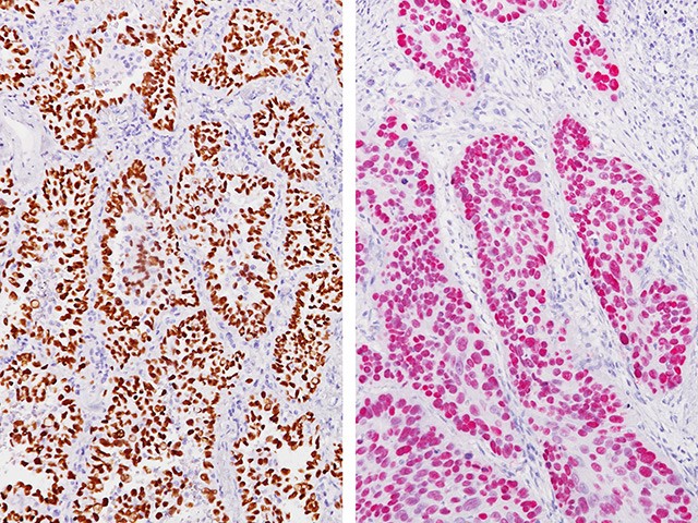
TTF-1 by IHC
TTF-1 by IHC-12376 - Technical only, 12379 - Technical & interpretation
LAB12379
- All IHC stains will include a positive control tissue
Applications
TTF-1 is present in normal thyroid and neoplastic thyroid tumors such as papillary carcinoma TTF-1 is present in the majority of pulmonary adenocarcinomas (73-76%) and pulmonary small cell carcinomas (83%) A smaller proportion of large cell undifferentiated lung carcinomas (26%) stain for TTF-1 Squamous cell carcinomas of the lung do not stain for TTF-1 Malignant mesotheliomas are negative for TTF-1 Cytoplasmic staining for TTF-1 can be useful in identifying hepatocellular carcinoma Papillary nasopharyngeal carcinomas have been reported to stain for this marker A small proportion of breast cancers will stain for this marker (2.4% of 546 primary breast cancers in one study) Small cell carcinomas of the prostate are positive for TTF-1 Colorectal adenocarcinoma (including metastatic to the lung) can show diffuse, moderate to strong TTF-1 positivity. It is estimated up to 3% of colorectal adenocarcinomas will be TTF-1 positive with the TTF-1 clone we use (Dako clone 8G7G3/1). While the majority of these cases will demonstrate weak TTF-1 positivity, diffuse, strong positivity can occur Other tumors have been rarely reported to be TTF-1 positive (nuclear), and include cholangiocarcinoma, endometrial, ovarian, gastric/esophageal, cervical, pancreatic ductal, urothelial, hepatocellular, adenoid cystic, other salivary gland carcinomas, and adrenal cortical carcinoma. Typically, these tumors will show expression in a small fraction of tumor cells
Submit a formalin-fixed, paraffin embedded tissue block
Formalin-fixed, paraffin embedded (FFPE) tissue block
FFPE tissue section mounted on a charged, unstained slide
Ambient (preferred)
- Unlabeled/mislabeled block
- Insufficient tissue
- Slides broken beyond repair
Immunohistochemical staining and microscopic examination
If requested, an interpretive report will be provided
Specifications
- Reacts with a transcription factor that is expressed selectively in the thyroid, lung, and diencephalon
- In the lung, TTF-1 is present in Type II Cells and Clara Cells and is important in the activation of pulmonary-specific differentiation genes
- In thyroid, TTF-1 is present in follicular epithelial cells
- TTF-1 is expressed early in the embryogenesis of thyroidal and respiratory epithelium, arising from endoderm of the foregut
- TTF-1 expression has also been found in some endometrial and endocervical carcinomas8
- Cytoplasmic staining for TTF-1 with this clone can be seen in hepatocellular carcinoma
Staining patterns
- Nuclear staining in lung, thyroid, and papillary nasopharyngeal carcinoma
- Cytoplasmic staining in hepatocellular carcinoma
88341 - each additional stain
