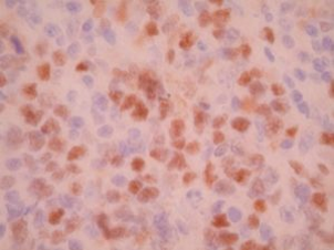
BCL-6 by IHC
BCL-6 by IHC-12376 - Technical only, 12379 - Technical & interpretation
BCL-6 by IHC
12376 - Technical only, 12379 - Technical & interpretation
LAB12376
LAB12379
LAB12379
All IHC stains will include a positive control tissue
- BCL6 staining is seen in follicular lymphomas, approximately 30% of diffuse large B cell lymphomas, Burkitt lymphomas, and nodular lymphocyte-predominant Hodgkin’s disease
- In normal tissues, BCL6 stains follicular center B-cells of the germinal centers, and some T-cells in the germinal centers; the staining pattern is reciprocal to that of Bcl-2
- BCL6 staining is not seen in B-CLL, hairy cell leukemia, and mantle and marginal-zone lymphomas
Tissue
Submit a formalin-fixed, paraffin embedded tissue block
Formalin-fixed, paraffin embedded (FFPE) tissue block
Tissue section mounted on a charged, unstained slide
Ambient (preferred)
- Unlabeled/mislabeled block
- Insufficient tissue
- Slides broken beyond repair
AHL - Immunohistochemistry
Mo - Fr
1 - 2 days
Immunohistochemical staining and microscopic examination
If requested, an interpretive report will be provided
Specifications
- BCL6 is a protein expressed by germinal center B-lymphocytes and serves as a transcriptional regulatory protein
- BCL6 expression is seen in a variety of B cell lymphomas
Staining patterns
- Nuclear staining
References
- Falini B, et al: Bcl-6 protein expression in normal and neoplastic lymphoid tissues. Ann Oncol 8, Suppl, 2:101-104, 1997.
- Flenghi L, et al: A specific monoclonal antibody detects expression of the BCL-6 protein in germinal center B cells. Am J Pathol 147:405-411, 1995.
- Falini B, et al: Distinctive expression pattern of the BCL-6 protein in nodular lymphocyte predominance Hodgkin’s disease. Blood 87:465-71, 1996.
88342 - 1st stain
88341 - each additional stain
88341 - each additional stain
05/10/2017
10/17/2018
01/12/2024
