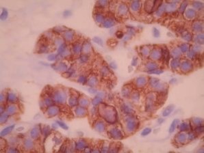
HBME-1 by IHC
HBME-1 by IHC-12376 - Technical only, 12379 - Technical & interpretation
HBME-1 by IHC
12376 - Technical only, 12379 - Technical & interpretation
LAB12376
LAB12379
LAB12379
HBME
- All IHC stains will include a positive control tissue
- Useful in identifying mesothelial cells (benign and malignant), although this marker cross-reacts with many tumors of other sites (such as from ovarian, lung, and GI tract)
- This marker is most useful in identifying papillary thyroid cancer; diffuse and intense membranous staining favors a papillary cancer
- Useful in battery with CK19 in discriminating papillary thyroid cancer from benign entities
- In a recent report 2, HMBE1 staining was observed in 87 % of papillary thyroid cancers, including 43 of 49 (88%) classic papillary thyroid cancers and 25 of 29 (86%) follicular variants of papillary thyroid cancer
- CK19 staining has been reported in 96% of papillary thyroid cancers, including 100% of classic papillary thyroid cancer and in 26 of 29 cases (90%) of follicular variants of papillary thyroid cancer 2
- The co-expression of HBME1 and CK19 has been reported as 100% specific and 83% sensitive 2
- HMBE1 staining may rarely be seen in follicular adenomas (2 of 49 cases; 4%); this staining pattern is usually focal, rarely diffuse and strong. CK19 has been seen in 14% (7 of 29) of follicular adenomas
- In one study, 73% of lung adenocarcinomas are positive for this marker
Tissue
Submit a formalin-fixed, paraffin-embedded tissue
Formalin-fixed, paraffin-embedded (FFPE) tissue block
FFPE tissue section mounted on a charged, unstained slide
Ambient (preferred)
- Unlabeled/mislabeled block
- Insufficient tissue
- Slides broken beyond repair
AHL - Immunohistochemistry
Mo - Fr
1 - 2 days
Immunohistochemical staining and microscopic examination
If requested, an interpretive report will be provided
Specifications
- HBME-1 reacts with an unknown antigen in the microvilli of mesothelial cells
- This antibody also reacts with a variety of benign and malignant epithelial cells
- HBME-1 will stain the majority of papillary thyroid cancers (including follicular variants); a negative result helps rule against a papillary cancer
- Non-specific staining for this marker can be seen in benign thyroid lesions and occasionally in follicular carcinomas
Staining pattern
- Membranous staining is most useful (cytoplasmic staining can also be seen)
References
- Miettinen M et al: HBME-1, a monoclonal antibody useful in the differential diagnosis of mesothelioma, adenocarcinoma and soft-tissue and bone tumors. Appl Immunohistochem 1995; 3(2):115.
- Scognamiglio T et al: Diagnostic usefulness of HBME1, Galactin-3, CK19, and CITED1 and evaluation of their expression in encapsulated lesions with questionable features of papillary thyroid carcinoma. Am J Clinc Pathol 2006; 126:700-708.
- Nasr M et al: Immunohistochemical markers in diagnosis of papillary thyroid carcinoma: utility of HBME1 combined
88342 - 1st stain
88341 - each additional stain
88341 - each additional stain
07/03/2017
10/19/2018
01/21/2026
