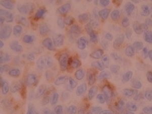
Tyrosinase by IHC
Tyrosinase by IHC-12376 - Technical only, 12379 - Technical & interpretation
Tyrosinase by IHC
12376 - Technical only, 12379 - Technical & interpretation
LAB12376
LAB12379
LAB12379
- All IHC stains will include a positive control tissue
- Highly specific marker for identifying melanoma
- Tyrosinase will not cross react with melanophages, which may obscure the identification of dermal melanocytic cells
- This marker is 100% specific and >80% sensitive for non-spindle cell type melanomas<
- Tyrosinase is 100% specific and 85% sensitive for amelanotic melanomas
- This marker can help identify cellular blue nevi that may be S100 negative
- Tyrosinase is a very specific marker for identifying malignant melanoma in sentinel lymph nodes
Tissue
Submit a formalin-fixed, paraffin embedded tissue block
Formalin-fixed, paraffin embedded (FFPE) tissue block
FFPE tissue section mounted on a charged, unstained slide
Ambient (preferred)
AHL - Immunohistochemistry
Mo - Fr
1 - 2 days
Immunohistochemical staining and microscopic examination
If requested, an interpretive report will be provided
Specifications
- Tyrosinase is an enzyme, that catalyzes the reaction that forms melanin
- This antigen is a highly specific marker for melanocytic differentiation, and is only present in melanin producing cells
Staining pattern
- Cytoplasmic granular staining (a red chromogen is used)
References
- Kaufmann O et al: Tyrosinase, melan-A, and KBA62 as markers for the immunohistochemical identification of metastatic amelanotic melanomas on paraffin sections. Mod Pathol 1998; 11:740-746.
- Orchard GE. Comparison of immunohistochemical labeling of melanocyte differentiation antibodies melan-A, tyrosinase, and HMB-45 with NK1/C3 and S100 protein in the evaluation of benign nevi and malignant melanoma. Histochem J 2000:32:475-481.
88342 - 1st stain
88341 - each additional stain
88341 - each additional stain
09/13/2017
10/17/2018
