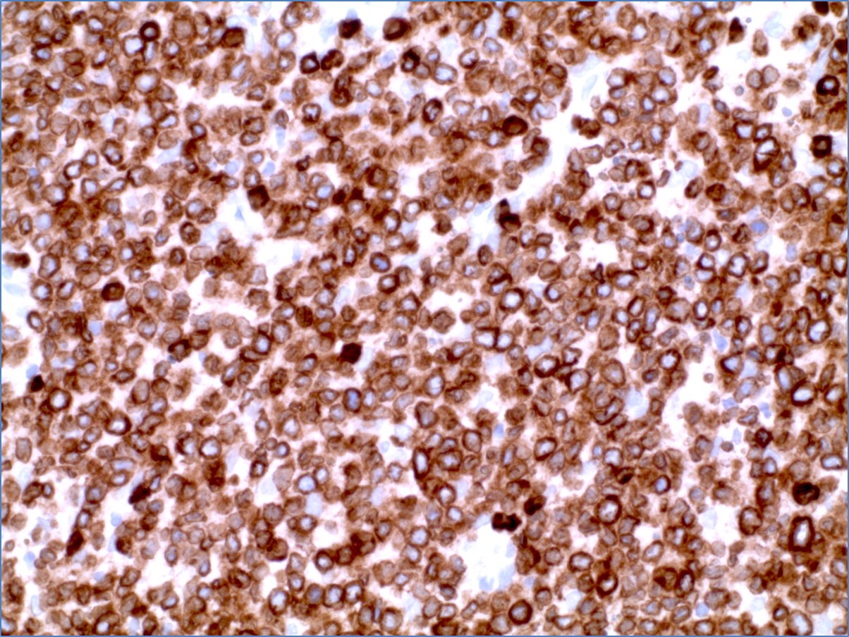
CD79a by IHC
CD79a by IHC-12376 - Technical only, 12379 - Technical & Interpretation
CD79a by IHC
12376 - Technical only, 12379 - Technical & Interpretation
LAB12376
LAB12379
LAB12379
pan B cell
All IHC stains will include a positive control tissue
- Intended for the identification of cells of B-cell lineage (reacts with 97% of B-cell neoplasms) [see Table 1]
- Staining is consistently strong on small cell neoplasm, but large B-cell neoplasms and follicular lymphomas show weaker staining2
- Normal plasma cells will stain intensely with CD79a
- 50% of myelomas show CD79a positivity2
- CD79a is found in the majority (89%) of acute leukemias of precursor B-cell type, in B-cell lines but not in T-cell lines1
- Most reliable B-cell marker detectable in paraffin embedded specimens in precursor B-cell ALL2
- CD79a is specifically associated with bi-phenotypic leukemias that express myeloid and B-cell antigens and have heavy chain genes in rearranged configurations. Along with CD-22, it is more useful than other B-cell antigens in detecting bi-phenotypic cases within AML
- CD79a is an improvement on L26(CD-20) which labels dendritic reticulum cells, some T-cells and reacts with neoplastic cells in cases of AML2
- CD20 is not expressed until the late precursors B-cell stage and at least half of precursor B ALL's are CD-20 negative3
- Occasional Reed Sternberg cells in cases of Hodgkin's disease are CD79a positive 4 & 5
Table 1 (Ref 2)
Reactivity of Anti-CD79a (mb-1) antibody JCB117 with paraffin-embedded samples of Hematopoietic Neoplasms
| B-cell neoplasms | |
|---|---|
| Small lymphocytic lymphoma/CLL | 28/28 |
| Lymphoplasmacytoid lymphoma | 36/36 |
| Mantle cell lymphoma | 17/17 |
| Follicular lymphoma | 53/53 |
| MALT lymphoma | 29/29 |
| Hairy cell leukemia | 15/15 |
| Myeloma/plasmacytoma | 10/20 |
| Large cell lymphoma | 95/95 |
| Burkitt's lymphoma | 7/7 |
| Anaplastic large cell lymphoma | 13/15 |
| Total | 344/356 (97%) |
| T-cell and non-lymphoid neoplasms | |
| Lymphoblastic lymphoma/leukemia | 0/9 |
| Mycosis fungoides | 0/10 |
| Peripheral T-cell lymphoma | 0/32 |
| Angioimmunoblastic T-cell lymphoma | 0/8 |
| Anaplastic large cell lymphoma | 0/11 |
| Acute myeloid leukemia | 0/28 |
| Total | 0/98 |
Tissue
Submit a formalin-fixed, paraffin-embedded tissue
Formalin-fixed, paraffin-embedded (FFPE) tissue block
FFPE tissue section mounted on a charged, unstained slide
Ambient (preferred)
- Unlabeled/mislabeled block
- Insufficient tissue
- Slides broken beyond repair
AHL - Immunohistochemistry
Mo - Fr
1 - 2 days
Immunohistochemical staining and microscopic examination
If requested, an interpretive report will be provided
Specifications
- The mb-1 polypeptide is part of the B-cell antigen receptor complex
- Appears early in B-cell maturation at the pre B-cell stage and persists until the plasma cell stage where it is found as an intracellular component
- Virtually B-cell specific
Staining pattern
- Cell surface membrane and cytoplasmic
References:
- Blood, Vol 82(3):853-857, 1993.
- Blood, Vol 86(4):1453-1459, 1995.
- AJCP, Vol 106:462-468, 1996.
- AJSP, Vol 19(11):1294-1299, 1995.
- Histopathology, 24(6):511-515, 1994.
- Spec sheet from DAKO.
88342 - 1st stain
88341 - each additional stain
88341 - each additional stain
06/16/2017
10/18/2018
01/12/2024