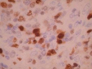
MUM1 by IHC
MUM1 by IHC-12376 - Technical only, 12379 - Technical & interpretation
MUM1 by IHC
12376 - Technical only, 12379 - Technical & interpretation
LAB12376
LAB12379
LAB12379
Multiple myeloma oncogene-1
- All IHC stains will include a positive control tissue
- MUM1 is useful in the subclassification of lymphoid malignancies
- MUM1 expression has been identified in multiple myelomas, lymphoplasmacytic lymphomas, diffuse large B-cell lymphomas, and activated T cells<
- MUM1 can also be used to identify Reed-Sternberg cells in Hodgkin's lymphoma
- MUM1 is also present in benign and neoplastic melanocytic proliferations (92% of melanomas are MUM1 positive)
- The presence of MUM1 staining in diffuse large B-cell lymphomas has been associated with a worse prognosis
Tissue
Submit a formalin-fixed, paraffin embedded tissue block
Formalin-fixed, paraffin embedded (FFPE) tissue block
FFPE tissue section mounted on a charged, unstained slide
Ambient (preferred)
- Unlabeled/mislabeled block
- Insufficient tissue
- Slides broken beyond repair
AHL - Immunohistochemistry
Mo - Fr
1 - 2 days
Immunohistochemical staining and microscopic examination
If requested, an interpretive report will be provided
Specifications
- Recognizes the MUM1/IRF4 protein, which is expressed in late plasma-cell-directed stages of B-cell differentiation
- MUM1 is useful in identifying transition from BCL6 positivity to CD138 expression
- It is also expressed in activated T cells and in related hematologic neoplasms
Staining pattern
- Nuclear staining, often with weak to moderate cytoplasmic staining
References:
- Colomo L et al: Diffuse large B-cell lymphomas with plasma-blastic differentiation represent a heterogeneous group of disease entities. Am J Surg Pathol. 2004 Jun;28(6): 736-47.
- Sundram U et al: Expression of the B-cell proliferation marker MUM1 by melanocytic lesions and comparison with S100, gp100 (HMB45), and MelanA. Mod Pathol. 2003 Aug;16(8):802-10.
- Camacho FI et al: Nodal marginal zone lymphoma: a heterogeneous tumor: a comprehensive analysis of a series of 27 cases. Am J Surg Pathol. 2003 Jun;27(6):762-71.
- Natkunam Y et al: Analysis of MUM1/IRF4 protein expression using tissue micro-arrays and immunohistochemistry. Mod Pathol. 2001 Jul;14(7):686-94.
- Chang CC et al: Immunohistochemical expression patterns of germinal center and activation B-cell markers correlate with prognosis in diffuse large B-cell lymphoma. Am J Surg Pathol. 2004 Apr;28(4):464-70.
- Hans CP et al: Confirmation of the molecular classification of diffuse large B-cell lymphoma by immunohistochemistry using a tissue micro-array. Blood. 2004 Jan 1;103(1):275-82. Epub 2003 Sep 22.
88342 - 1st stain
88341 - each additional stain
88341 - each additional stain
07/18/2017
10/19/2018
01/12/2024