MOC-31 by IHC
MOC-31 by IHC-12376 - Technical only, 12379 - Technical & interpretation
MOC-31 by IHC
12376 - Technical only, 12379 - Technical & interpretation
LAB12376
LAB12379
LAB12379
- All IHC stains will include a positive control tissue
- The primary use for this antibody is in distinguishing between epithelial versus mesothelial proliferations. It is one of the best markers currently available in recognizing adenocarcinoma
- This antibody should be used in a panel (see mesothelioma vs. adenocarcinoma panel)
Tissue
Submit a formalin-fixed, paraffin embedded tissue block
Formalin-fixed, paraffin embedded (FFPE) tissue block
FFPE tissue section mounted on a charged, unstained slide
Ambient (preferred)
- Unlabeled/mislabeled block
- Insufficient tissue
- Slides broken beyond repair
AHL - Immunohistochemistry
Mo - Fr
1 - 2 days
Immunohistochemical staining and microscopic examination
If requested, an interpretive report will be provided
Specifications
- MOC-31 is an antibody directed against a small cell carcinoma cell line
- It reacts with an epithelial antigen present on most normal and malignant epithelia
- Reactive mesothelial cells and malignant mesotheliomas show rare expression with this antibody
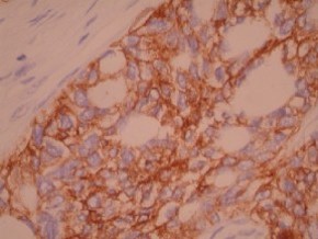
Staining pattern
- Membrane based staining
References
- Sosolik RC, et al. Anti-MOC-31: a potential addition to the pulmonary adenocarcinoma versus mesothelioma immunohistochemistry panel. Mod Pathol 1997; 10:716-719.
- Ordonez NG. The immunohistochemical diagnosis of epithelial mesothelioma. Hum Pathol 1999; 30:313-23.
- Ruitenbeek T, et al. Immunocytology of body cavity fluids. MOC031, a monoclonal antibody discriminating between mesothelial and epithelial cells. Arch Pathol Lab Med 1994; 118:265.
88342 - 1st stain
88341 - each additional stain
88341 - each additional stain
07/17/2017
10/19/2018
01/12/2024