PHH3 by IHC
PHH3 by IHC-12376 - Technical only, 12379 - Technical & interpretation
PHH3 by IHC
12376 - Technical only, 12379 - Technical & interpretation
LAB12376
LAB12379
LAB12379
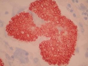
PHH3
Phosphohistone
Phosphohistone
- All IHC stains will include a positive control tissue
- PHH3 is an effective marker of mitotic figures
- It can be used to identify mitoses and help classify tumor grades, such as in tumors in CNS, skin, gynecologic, soft tissue and GIST
- In contrast, Ki67 is expressed in cells throughout the cell cycle from late G1 phase (except G0); and since many cells do not survive the cell cycle, the Ki67 proliferation index may not be representative of the proliferative potential of a tumor
- This antibody has been shown to be a helpful adjunct in classifying meningiomas1, 5, and a recent study suggests it may be used to help predict survival4
- PHH3 may help in the classification of astrocytomas2
- Studies have shown that PHH3 expression is higher in malignant melanomas as compared to Spitz nevi, and thus both PHH3 and Ki67 can be used in separating malignant melanomas from benign nevi3
Tissue
Prepare a formalin-fixed, paraffin-embedded (FFPE) tissue block
Formalin-fixed, paraffin-embedded (FFPE) tissue block
FFPE tissue section
Mount FFPE tissue section on a charged, unstained slide

Ambient (preferred)
- Unlabeled/mislabeled block
- Insufficient tissue
- Slides broken beyond repair
AHL - Immunohistochemistry
Mo - Fr
1 - 2 days
Immunohistochemical staining and microscopic examination
If requested, an interpretive report will be provided
Specifications
- PHH3 is a histone protein is present during chromatin condensation during mitosis (and it is not present during apoptosis)
- This antibody is useful to label mitoses (and separate from apoptotic bodies and karyorrhectic debris)
Staining pattern
- Nuclear
References
- Kim YJ et al: Prognostic significance of the mitotic index using the mitosis marker anti-phospho-histone H3 in meningiomas. Am J Clin Pathol. 2007 Jul;128(1):118-25.
- Coleman H et al: Assessment and prognostic significance of mitotic index using the mitosis marker phospho-histone H3 in low and intermediate-grade infiltrating astrocytomas. Am J Surg Pathol. 2006 May;30(5):657-64.
- Nasr MR et al. Comparison of pHH3, Ki-67, and survivin immunoreactivity in benign and malignant melanocytic lesions. Am J Dermatopathol. 2008 Apr;30(2):117-22. doi: 10.1097/DAD.0b013e3181624054.
- Olar A et al: Mitotic Index is an Independent Predictor of Recurrence-Free Survival in Meningioma. Brain Pathol. 2014 Jul 18. doi: 10.1111/bpa.12174.
- Ribalta T et al: The mitosis-specific antibody anti-phosphohistone-H3 (PHH3) facilitates rapid reliable grading of meningiomas according to WHO 2000 criteria. Am J Surg Pathol. 2004 Nov;28(11):1532-6.
88342 - 1st stain
88341 - each additional stain
88341 - each additional stain
08/07/2017
01/31/2024
01/31/2022