ROS1 by IHC
ROS1 by IHC-12379 - Technical & interpretation
- All IHC stains will include a positive control tissue
The ROS1 IHC test is a screening test. All cases considered to be positive for ROS1 IHC must be confirmed by ROS1 FISH testing in order to determine eligibility for crizotinib therapy.
Submit a formalin-fixed, parafin embedded (FFPE) tissue block
Formalin-fixed, paraffin embedded (FFPE) tissue block
FFPE tissue section mounted on a charged, unstained slide
Ambient (preferred)
Immunohistochemical staining and microscopic examination
Antibody
- Cell Signaling Technology (clone D4D6)
- Rabbit monoclonal antibody
- RUO
Specifications
- The incidence of ROS1 gene rearrangements in lung adenocarcinomas is estimated to range from 1-2%
- ROS1 is a tyrosine kinase and is an insulin receptor
- The function of ROS1 is undefined, but known to be involved in differentiation of epididymal epithelium
- ROS1 fusion gene rearrangements have been identified in glioblastoma, ovarian cancer, and in lung adenocarcinoma and lung non-small cell carcinoma NOS
Staining pattern
- Membranous, cytoplasmic globular, or finely cytoplasmic staining.
- The corresponding H&E stained section should be reviewed concurrently with the ROS1 IHC stained slide.
Negative staining
Lack of staining in tumor cells. The literature recommends at least 20 tumor cells be evaluated before calling a case negative.
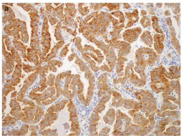
Examples of ROS1 IHC staining patterns 2
a) Diffuse intracytoplasmic globular reactivity
b) Focal intracytoplasmic globular reactivity
c) Reactivity localized to the lateral surface
d) Reactivity localized along the apical surface
Diffuse (a) or focal (b) instracytoplasmic globular reactivity was observed in 6 of 10 CD74-ROS1-positive cancers. Plasma membranous accentuation with a fine granular quality was observed in 3 of 4 EZR-ROS1-positive tumors; reactivity localized to the lateral surface in two cases (c) and along the apical surface in one case (d).
EZR-ROS1 positive signet-ring cell carcinoma (a) showed diffuse but only weak-moderate ROS1 immunoreactivity.
Known stain artifacts
- Weak positive staining can be present in alveolar macrophages, reactive type II pneumocyte hyperplasia, and bronchial metaplasia
- In bone metastasis, osteoclasts can demonstrate strong positivity
- False negative ROS1 IHC results have been reported in signet ring lung adenocarcinomas
- The majority of invasive mucinous lung adenocarcinomas will show positivity despite lacking the ROS1 gene rearrangement
- Staining of unknown specificity has been observed in cholangiocarcinoma, hepatocellular carcinoma and kidney
References
- Bubendorf L, Buttner R, Al-Dayel F, et al. Testing for ROS1 in non-small cell lung cancer: a review with recommendations. Virchows Arch 2016;469:489-503.
- Yoshida A, Tsuta K, Wakai S. Immunohistochemical detection of ROS1 is useful for identifying ROS1 rearrangements in lung cancers. Modern Pathology 2014;27:711-720
88341 - each additional stain