MiTF by IHC
MiTF by IHC-12376 - Technical only, 12379 - Technical & Interpretation
MiTF by IHC
12376 - Technical only, 12379 - Technical & Interpretation
LAB12376
LAB12379
LAB12379
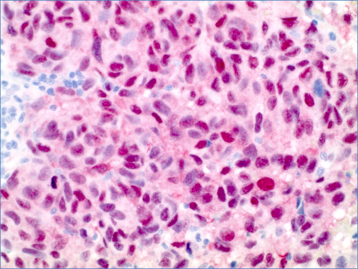
Microphthalmia transcription factor
- All IHC stains will include a positive control tissue
- Anti MITF recognizes a nuclear protein which is expressed in most primary and metastatic epithelioid malignant melanomas
- MITF is also seen in normal melanocytes, benign nevi and dysplastic nevi
- This marker is present in a small subset of non-melanoma tissues and tumors (heart, macrophages, rare cells of pituitary, endometrial stromal cells, breast cancer, leiomyosarcoma, malignant mixed mullerian tumors, and renal cell carcinomas
Tissue
Submit a formalin-fixed, paraffin-embedded (FFPE) tissue block
FFPE tissue block
FFPE tissue section mounted on a charged, unstained slide
Ambient (preferred)
AHL - Immunohistochemistry
Mo - Fr
1 - 2 days
Immunohistochemical staining and microscopic examination
If requested, an interpretive report will be provided
Specifications
- MITF is a transcription factor that regulates the development and survival of melanocytes and retinal pigment epithelium
- It is also involved in the to enzyme genes such as tyrosinase, TRP1 and TRP2
Staining pattern
- Nuclear
References
- Chang KL et al: Diagnostic utility of microphthalmia transcription factor in malignant melanoma and other tumors. Adv Anat Pathol. 2001; 8(5):273.
- Granter SR et al: Rogle for MITF in the diagnosis of metastatic malignant melanoma. Appl Immunhistochem Mol Morphol 2002; 10(1):47.
88342 - 1st stain
88341 - each additional stain
88341 - each additional stain
07/17/2017
10/19/2018
10/02/2020