p16 by IHC
p16 by IHC-12376 - Technical only, 12379 - Technical & Interpretation
p16 by IHC
12376 - Technical only, 12379 - Technical & Interpretation
LAB12376
LAB12379
LAB12379
Ink4
- All IHC stains will include a positive control tissue
- P16 is a marker for high risk HPV; useful in identifying SIL associated with high-risk HPV types
- Particularly useful in determining whether atypical squamous metaplasia is HSIL
- Staining for p16 correlates with a positive predictive value for HPV (88.7%)
- 95.8% of LSIL and 92% of HSIL stain for p16 (low risk strains of HPV-related neoplasia are less likely to stain as compared to high risk strains)
- Note: HPV negative carcinomas can also stain for this, indicating different pathways leading to p16 over-expression
- P16 will stain tubal metaplasia
- p16 will identify cervical adenocarcinoma, particularly those associated with HPV 16/18 infections
- Normal epithelium is p16 negative; inflammatory and metaplastic lesions are p16 negative or will show just focal staining
Tissue
Submit a formalin-fixed, paraffin embedded tissue block
Formalin-fixed, paraffin embedded (FFPE) tissue block
FFPE tissue section mounted on a charged, unstained slide
Ambient (preferred)
- Unlabeled/mislabeled block
- Insufficient tissue
- Slides broken beyond repair
AHL - Immunohistochemistry
Mo - Fr
1 - 2 days
Immunohistochemical staining and microscopic examination
If requested, an interpretive report will be provided
Specifications
- p16 (ink4) is a cell cycle associated protein which inhibits cyclin-dependent kinases by blocking substrate phosphorylation of the tumor suppressor retinoblastoma protein (Rb) and Rb related proteins p107 and p130 (thus interfering with the cell cycle)
- The gene that codes for p16 (CDKN2 gene) is mutated in a high percentage of tumors (including 75% of melanomas, as well as other cancers including breast and pancreas)
- The functional inactivation of p16 (as by HPV related infections in cervical lesions) is thought to lead to over-expression of this protein (and loss of its cell-cycle inhibitory effect)
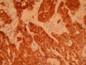
Staining pattern
- Cytoplasmic, with some nuclear staining also observed
- Staining in cervical neoplasia has been graded as1:
- Positive - continuous staining the horizontal plane, either partial or full thickness
- Focal positive - less than 80% staining in an interrupted plane
- Negative - weakly diffuse staining characterized by a "blush"
References
- Keating JT et al: Ki-67, Cyclin E and p16 are complimentary surrogate bio-markers for HPV-related cervical neoplasia. Am J Surg Pathol 25(7): 884-891, 2001.
- Sano T et al: Expression status of p16 protein is associated with HPV oncogenic potential in cervical and genital lesions. Am J Pathol 1998; 153:1741-8, 1998.
- Hirama T et al: p16 gene is not altered in uterine cervical carcinomas or cell lines. Mod Pathol 1996; 9:26-31.
- Milde-Langosch K et al: Expression of cyclin-dependent kinase inhibitors p16 etc. in HPV positive and HPV negative cervical adenocarcinoma. Virchows Arch (2001) 439; 55-61.
- Klaes R et al: Over-expression of p16 as a specific marker for dysplastic and neoplastic epithelial cells of the cervix uteri. Int J Cancer: 92, 276-284 (2001).
- Keating JT et al: Surrogate bio-markers of HPV infection in cervical neoplasia screening and diagnosis. Adv Anat Pathol, vol 8, no 2, 83-92, 2001.
- Negri G, et al: P16 is a useful marker for the diagnosis of adenocarcinoma of the cervix uteri and its precursors; an immunohistochemical study with immunocytochemical correlations, Am J Surg Pathol 27 (2): 187-193, 2003.
- Kong CS et al: p16 immunohistochemistry is superior to HPV in situ hybridization for the detection of high-risk PHPV in atypical squamous metaplasia. Am J Surg Pathol 2007; 31:33-43).
88342 - 1st stain
88341 - each additional stain
88341 - each additional stain
07/18/2017
10/19/2018
01/12/2024