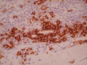
CD138 by IHC
CD138 by IHC-12376 - Technical only, 12379 - Technical & interpretation
CD138 by IHC
12376 - Technical only, 12379 - Technical & interpretation
LAB12376
LAB12379
LAB12379
Syndecan-1
- All IHC stains will include a positive control tissue
- CD138 is a very useful marker in identifying plasma cells
- This antibody will identify plasmacytomas/multiple myeloma, plasmocytic lymphomas, and some cases of immunoblastic lymphoma with plasmacytoid differentiation
- CD138 can be used in a panel with bcl-6, to help divide the HIV associated lymphomas into those of follicular origin (bcl-6 + / CD138 -) and those of post-follicular origin (bcl-6 - / CD138 +)
- CD138 staining has been shown to be present in normal epidermis, squamous cell carcinoma in situ, and keratoacanthomas; in contrast, invasive squamous cell carcinomas show markedly reduced staining
CD138 reactivity in plasmacytic and non-plasmacytic tumors7
| Diagnosis | # cases | Tumor cells | Stroma |
| Multiple myeloma | 43 | 43 | 7 |
| Lymphoplasmacytic NHL | 4 | 4 | 0 |
| Breast cancer | 9 | 8 | 7 |
| Gastric cancer | 5 | 4 | 1 |
| Small cell lung cancer | 2 | 1 | 2 |
| Non-small cell lung cancer | 5 | 5 | 5 |
| Colon cancer | 3 | 3 | 1 |
| Hepatocellular cancer | 2 | 2 | 0 |
| Renal cell cancer | 1 | 1 | 0 |
| TCC bladder | 3 | 3 | 0 |
| Papillary thyroid | 1 | 1 | 0 |
| Myoepithelioma, salv. gland | 1 | 1 | 0 |
| Thymoma | 1 | 1 | 0 |
| Mesothelioma | 2 | 2 | 0 |
| Pheochromocytoma | 2 | 0 | 0 |
| Melanoma | 10 | 5 | 6 |
| Seminoma | 1 | 0 | 0 |
| Synovial sarcoma | 2 | 2 | 0 |
| GIST | 1 | 1 | 0 |
| Schwannoma | 1 | 0 | 0 |
| Leiomyosarcoma | 2 | 1 | 0 |
Tissue
Submit a formalin-fixed, paraffin embedded tissue block
Formalin-fixed, paraffin embedded (FFPE) tissue block
FFPE tissue section mounted on a charged, unstained slide
Ambient (preferred)
- Unlabeled/mislabeled block
- Insufficient tissue
- Slides broken beyond repair
AHL - Immunohistochemistry
Mo - Fr
1 - 2 days
Immunohistochemical staining and microscopic examination
If requested, an interpretive report will be provided
Specifications
- CD138 is a plasma cell-associated transmembrane heparan sulfate proteoglycan, that mediates cell to matrix adhesion; its expression is inversely correlated with tumor aggressiveness and invasiveness
- CD138 is expressed in plasma cells, as well as pre-B cells, post-follicular immunoblasts
- CD138 can react with Reed-Sternberg cells (classical Hodgkin's disease, results vary from high degree of staining in some studies, to only stromal staining in other studies)
- This antibody is not specific for plasmacytic derivation, since it will also stain epithelial cells (keratinocytes, and simple and stratified epithelial cells), as well as stromal cells and melanocytic cells (see below)
- CD138 expression is not definitive for plasmacytic derivation, unless a hematolymphoid phenotype has been established, and a possible epithelial or stromal (including melanocytic) process has been excluded
Staining pattern
- Cell surface membrane staining
References
- King BE et al: Immunophenotypic and genotypic markers of follicular center cell neoplasia in diffuse large B-cell lymphomas. Mod Pathol 2000; 13(11):1219-1231.
- Capello D et al: Molecular pathophysiology of indolent lymphoma. Haematologica 2000; 183:195-201.
- Bayer-Garner IB et al: Syndecan-1 (CD138) immunoreactivity in bone marrow biopsies of multiple myeloma: shed syndecan-1 accumulates in fibrotic regions. Mod Pathol 2001 Oct; 14(10):1052-8.
- Chilosi M et al: CD138/syndecan-1: a useful immunohistochemical marker of normal and neoplastic plasma cells on routine trephine bone marrow biopsies. Mod Pathol 1999 Dec; 12(12):1101-6.
- Costes V et al: The Mi15 monoclonal antibody (anti-syndecan-1) is a reliable marker for quantifying plasma cells in paraffin-embedded bone marrow biopsy specimens. Hum Pathol 1999 Dec; 30 (12):1405-11.
- Mukunyadzi P et al: The level of syndecan-1 expression is a distinguishing feature in behavior between keratoacanthoma and invasive cutaneous squamous cell carcinoma. Mod Pathol 2002 Jan; 15(1):45-9.
- O'Connell FP et al: CD138 (syndecan-1), a plasma cell marker; immunohistochemical profile in hematopoietic and non-hematopoietic neoplasms. Am J Clin Pathol 2004; 121:254-263
88342 - 1st stain
88341 - each additional stain
88341 - each additional stain
06/19/2017
10/17/2018
01/12/2024