NUT by IHC
NUT by IHC-12376 - Technical only, 12379 - Technical & Interpretation
NUT by IHC
12376 - Technical only, 12379 - Technical & Interpretation
LAB12376
LAB12379
LAB12379
IHC
- All IHC stains will include a positive control tissue
Tissue
Submit a formalin-fixed, paraffin embedded tissue block
Formalin-fixed, paraffin embedded (FFPE) tissue block
FFPE tissue section mounted on a charged, unstained slide
Ambient (preferred)
- Unlabeled/mislabeled block
- Insufficient tissue
- Slides broken beyond repair
AHL - Immunohistochemistry
Mo - Fr
1 - 2 days
Immunohistochemical staining and microscopic examination
If requested, an interpretive report will be provided
Specifications
- NUT gene (NUTM1, NUT midline carcinoma family member 1) on chromosome 15 encodes NUT protein (nuclear protein in testis, FAM22H and NUT family member 1). Function of NUT protein is uncertain.
- NUT carcinoma is a highly aggressive malignancy defined by translocations involving the NUT gene. Fusion with partner genes (BRD4, BRD3, NSD3, and others) leads to the deregulated gene transcription of multiple genes including cMYC and TP63.
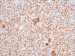
Staining pattern
Nuclear staining. Cytoplasmic staining is nonspecific.
- NUT carcinoma: Strong speckled nuclear staining in greater than 50% of tumor cells. Most NUT carcinomas demonstrate strong speckled staining in >90% of tumor cells.
- Majority of malignant ovarian germ cell tumors and a subset of other germ cell tumors (seminomas, embryonal carcinoma, and others) demonstrate nuclear staining in a subset of tumor cells. The nuclear staining is uniform (non-speckled) and has been reported to be weak to moderate in intensity.
- Staining in normal tissues: Germ cells of the testis and ovary demonstrate homogenous (non-speckled) nuclear staining.
88342 - 1st stain
88341 - each additional stain
88341 - each additional stain
07/30/2019
07/30/2019
01/12/2024