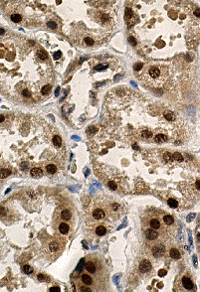
HNF-1 beta by IHC
HNF-1 beta by IHC-12376 - Technical only, 12379 - Technical & interpretation
HNF-1 beta by IHC
12376 - Technical only, 12379 - Technical & interpretation
LAB12376
LAB12379
LAB12379
Hepatocyte nuclear factor 1b
HNF-1b
HNF-1b
- All IHC stains will include a positive control tissue
HNF-1 should be used in a panel of immunohistochemical stains to differentiate clear cell carcinomas of the ovary and endometrium from other malignancies. At this time, HNF-1 is not considered to be sensitive or specific enough to use alone 1
- HNF-1 typically shows diffuse positivity in clear cell carcinomas of the ovary and endometrium (reported sensitivity of 82-89% and specificity of 55.9-97%)1-3
- HNF-1 positivity has been reported in: High grade serous carcinoma, endometrioid carcinoma of the uterus and ovary, yolk sac tumor, mesonephric adenocarcinoma, and carcinomas from the lung, thyroid, pancreas, liver, gastrointestinal tract and GU tract
- HNF-1 is positive in normal liver, pancreas, kidney, upper and lower GI tract, secretory endometrium, and gestational endometrium
Tissue
Submit a formalin-fixed, paraffin-embedded tissue
Formalin-fixed, paraffin-embedded (FFPE) tissue block
FFPE tissue section mounted on a charged, unstained slide
Ambient (preferred)
- Unlabeled/mislabeled block
- Insufficient tissue
- Slides broken beyond repair
AHL - Immunohistochemistry
Mo - Fr
1 - 2 days
Immunohistochemical staining and microscopic examination
If requested, an interpretive report will be provided
Specifications
- Component of the hepatocyte nuclear factor family, transcription factors which regulate glucose metabolism in liver, kidney, small intestine and thymus
Staining pattern
- Nuclear staining pattern; cytoplasmic staining is non-specific
References
- Kobel et al. A limited panel of immunomarkers can reliable distinguish between clear cell and high-grade serous carcinoma of the ovary. Am J Surg Pathol 2009;1(33):14-21.
- Lim et al. Immunohistochemical comparison of ovarian and uterine endometrioid carcinoma, endometrioid carcinoma with clear cell change and clear cell carcinoma. Am J Surg Pathol 2015;39(8):1061-1068.
- DeLair et al. Morphologic spectrum of immunohistochemically characterized clear cell carcinoma of the ovary: a study of 155 cases. Am J Surg Pathol 2011;35(1):36-44.
88342 - 1st stain
88341 - each additional stain
88341 - each additional stain
07/03/2017
10/19/2018
01/12/2024