Estrogen receptor by IHC
Estrogen receptor by IHC-12376 - Technical only, 12379 - Technical & interpretation
Estrogen receptor by IHC
12376 - Technical only, 12379 - Technical & interpretation
LAB12376
LAB12379
LAB12379
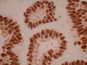
ER
All IHC stains will include a positive control tissue
- ER is used to determine if a tumor expresses estrogen receptor (i.e. for classification purposes)
- ER positivity is used to determine if a patient would be a candidate for anti-estrogen therapy
- Note: ER positivity may be seen in 5-10% of lung, nonsmall cell carcinomas (up to 18% with our previous antibody 1D5); staining for ER in lung tumors is usually focal and variable in intensity 7; In the context of an ER-positive lung neoplasm, strong and extensive TTF-1 immunoreactivity can be regarded as strong supportive evidence for a primary bronchogenic adenocarcinoma
Tissue
Submit a formalin-fixed, paraffin-embedded tissue
Formalin-fixed, paraffin-embedded (FFPE) tissue block
FFPE tissue section mounted on a charged, unstained slide
Ambient (preferred)
- Unlabeled/mislabeled block
- Insufficient tissue
- Slides broken beyond repair
AHL - Immunohistochemistry
Mo - Fr
1 - 2 days
Immunohistochemical staining and microscopic examination
If requested, an interpretive report will be provided
Specifications
- ER is a nuclear protein that binds estrogen hormone
- ER expression is seen in a variety of tumors from breast or gynecologic origin
- ER expression can also be seen in tumors of lung, stomach, thyroid, and very rarely colorectal origin
Staining pattern
- Nuclear staining
References
- Deamant FD, et al: Estrogen receptor immunohistochemistry as a predictor of site of origin in metastatic breast cancer. Appl Immunohistochem 1(3): 188-192, 1993.
- Esteban JM, et al: Biologic significance of quantitative estrogen receptor immunohistochemical assay by image analysis in breast cancer. Am J Clin Pathol 1994: 102:158-162.
- Aziz DC: Quantitation of estrogen and progesterone receptors by immunocytochemical and image analyses. Am J Clin Pathol 1992; 98:105-111.
- Wilbur DC et al: Estrogen and progesterone receptor detection in archival formalin-fixed, paraffin-embedded tissue from breast carcinoma: A comparison of immunohistochemistry with the dextran-coated charcoal assay. Mod Pathol, 5(1), 1992.
- Esteban JM et al: Improvement of the quantification of estrogen and progesterone receptors in paraffin-embedded tumors by image analysis. Am J Clin Pathol, 1993; 99:32-38.
- Taylor CR: Paraffin section immunocytochemistry for estrogen receptor; the time has come. Cancer 77(12), 1996.
- Lau SK et al: Immunohistochemical expression of estrogen receptor in pulmonary adenocarcinoma. Appl Immunohistochem Mol Morphol. 2006 Mar; 14 (1): 83-7.
88342 - 1st stain
88341 - each additional stain
88341 - each additional stain
06/21/2017
10/17/2018
01/12/2024